Posts By :

CACRelatyv

Relatyv - Everything to Know

Everything To Know About Collateral Ligament Pain In The Knee

Everything To Know About Collateral Ligament Pain In The Knee 2560 1442 Relatyv

Understanding Collateral Ligament Knee Pain

The collateral ligaments are essential stabilizers of the knee, helping control side-to-side movement and keeping the joint aligned during walking, running, jumping, and other daily activities. When these ligaments become stretched or torn, they can cause pain, swelling, instability, and difficulty moving the knee.

This guide explains the symptoms, causes, diagnosis, treatment options, and how our Relatyv Neurofunctional Pain Management approach may complement your overall treatment plan.

What Are the Collateral Ligaments?

The knee contains four major ligaments that stabilize the joint. Two of these are the collateral ligaments:

  • Medial Collateral Ligament (MCL): Located on the inside of the knee, the MCL connects the femur (thigh bone) to the tibia (shin bone) and helps prevent excessive inward movement.
  • Lateral Collateral Ligament (LCL): Located on the outside of the knee, the LCL connects the femur to the fibula and helps stabilize the outer portion of the knee.

These ligaments work together with the cruciate ligaments, muscles, tendons, and cartilage to provide stability during movement.

Symptoms of Collateral Ligament Injuries

Collateral ligament injuries commonly occur after a direct blow to the knee, twisting injury, or sudden change in direction. Symptoms may include:

  • Pain along the inside or outside of the knee.
  • Swelling.
  • Tenderness when touching the injured area.
  • A feeling that the knee is unstable or may “give way.”
  • Difficulty bending or straightening the knee.
  • Clicking, locking, or catching sensations.
  • Reduced range of motion.

Medial Collateral Ligament (MCL) Injuries

MCL injuries are among the most common ligament injuries of the knee. They often occur when a force strikes the outside of the knee, stretching the ligament on the inside.

Common symptoms include:

  • Pain along the inner knee.
  • Localized swelling.
  • Tenderness.
  • Difficulty fully bending or straightening the knee.

Lateral Collateral Ligament (LCL) Injuries

LCL injuries typically occur when force is applied to the inside of the knee, placing stress on the outer ligament.

Symptoms may include:

  • Pain along the outside of the knee.
  • Swelling and tenderness.
  • Instability while walking.
  • Difficulty bearing weight.

Common Causes of Collateral Ligament Pain

Sports Injuries

Contact sports and activities involving rapid changes in direction, jumping, or pivoting place significant stress on the collateral ligaments. Football, soccer, skiing, basketball, and hockey are common examples.

Sprains

A ligament sprain occurs when the ligament is stretched beyond its normal range. Depending on the severity, small or partial tears may develop.

Ligament Tears

More severe injuries may involve partial or complete tears of the ligament, leading to pain, swelling, and knee instability.

Degenerative Conditions

Osteoarthritis and other chronic joint conditions may alter knee mechanics and contribute to ligament stress over time.

How Collateral Ligament Injuries Are Diagnosed

Your healthcare provider may recommend:

  • Medical history review.
  • Physical examination to assess stability and range of motion.
  • X-rays to evaluate for fractures or bone injuries.
  • MRI to assess ligament damage and other soft tissues.
  • Arthroscopy in selected cases when additional evaluation is needed.

Grading Collateral Ligament Injuries

Grade 1

A mild sprain involving microscopic tearing of the ligament. The knee remains stable, and symptoms often improve with conservative treatment.

Grade 2

A partial tear causing moderate pain, swelling, tenderness, and some instability.

Grade 3

A complete tear of the ligament resulting in significant instability and loss of function. Surgical evaluation may be appropriate depending on the injury and individual needs.

Conventional Treatment Options

Treatment depends on the severity of the injury and your activity level.

Rest, Ice, Compression, and Elevation (RICE)

Mild injuries often improve with activity modification, icing, compression, and elevation during the early stages of recovery.

Bracing

A knee brace may provide additional stability while the ligament heals.

Physical Therapy

Rehabilitation exercises help restore strength, flexibility, balance, and normal movement patterns while supporting recovery.

Medications

NSAIDs or other pain-relieving medications may help reduce pain and inflammation. Your healthcare provider can discuss the benefits and potential risks based on your individual situation.

Surgery

Complete ligament tears or complex knee injuries may require surgical reconstruction when conservative treatment is not sufficient.

Our Relatyv Neurofunctional Pain Management Approach

At Relatyv, our Neurofunctional Pain Management approach combines drug-free, non-invasive therapies designed to help reduce chronic pain, improve function, and support healthy nervous system activity.

Each treatment plan is individualized based on your diagnosis, symptoms, and goals. Care may include electroanalgesia, IV therapy when clinically appropriate, nutritional support, and lifestyle recommendations that complement your existing medical care.

Address Knee Ligament Pain Early

Persistent knee pain, instability, or swelling should not be ignored. Early evaluation can help determine the extent of a ligament injury and guide the most appropriate treatment plan, reducing the risk of ongoing instability or additional knee damage.

At Relatyv, we provide personalized, non-invasive care designed to help reduce chronic pain, improve function, and support long-term mobility. Our Neurofunctional Pain Management approach may help complement your existing treatment plan while supporting your recovery and overall quality of life.


Relatyv - Crepitus

Crepitus: What Does The Grating Sensation Indicate?

Crepitus: What Does The Grating Sensation Indicate? 2394 1348 Relatyv

Understanding Joint Crepitus

Crepitus is the medical term for a grinding, cracking, popping, or crunching sensation that occurs when a joint moves. While occasional joint noises are common and often harmless, persistent crepitus—especially when accompanied by pain, swelling, or stiffness—may indicate an underlying joint or musculoskeletal condition that should be evaluated by a healthcare provider.

This guide explains what crepitus is, common causes, associated conditions, available treatment options, and how our Relatyv Neurofunctional Pain Management approach may complement your overall treatment plan.

How Healthy Joints Function

Joints are where two or more bones meet, allowing the body to move while providing stability and absorbing impact. Healthy joints contain several important structures that work together, including:

  • Cartilage to cushion the ends of bones.
  • Ligaments to stabilize the joint.
  • Tendons that connect muscles to bones and produce movement.
  • Joint capsule and synovial fluid that help lubricate and protect the joint.

When any of these structures become irritated, inflamed, injured, or worn over time, joint movement may produce sounds or sensations known as crepitus.

What Is Crepitus?

Crepitus refers to grinding, crackling, clicking, or popping sensations that occur during joint movement. Some people experience only the sound, while others also notice pain, stiffness, swelling, or reduced mobility.

Crepitus may occur in many joints, including the:

  • Knees
  • Shoulders
  • Hips
  • Ankles
  • Wrists
  • Elbows
  • Neck

Types of Crepitus

Joint Crepitus

This occurs when joint surfaces move against one another, often because of cartilage changes, inflammation, or altered joint mechanics.

Bone Crepitus

Bone crepitus may occur when fractured bone fragments move against one another.

Bursal Crepitus

Inflammation of the bursa (bursitis) may occasionally produce a grinding sensation during movement.

Tendon Crepitus

Inflamed tendons or tendon sheaths, such as with tenosynovitis, may create a crackling or rubbing sensation as the tendon moves.

Symptoms That May Occur with Crepitus

Depending on the underlying condition, crepitus may occur alongside:

  • Joint pain.
  • Swelling.
  • Stiffness.
  • Tenderness.
  • Warmth around the joint.
  • Reduced range of motion.

Conditions Associated with Crepitus

Osteoarthritis

As cartilage gradually wears away, bones may move less smoothly within the joint, contributing to pain, stiffness, and joint noises.

Rheumatoid Arthritis

Inflammation caused by rheumatoid arthritis may damage joint tissues over time, resulting in pain, swelling, stiffness, and crepitus.

Gout

Uric acid crystal deposits within the joints may cause painful inflammation and contribute to abnormal joint movement.

Fibromyalgia

Some individuals with fibromyalgia report sensations of joint or muscle crepitus alongside chronic widespread pain and stiffness.

Lyme Disease

Lyme disease may cause inflammatory arthritis affecting one or more joints, particularly the knees.

Systemic Lupus Erythematosus (SLE)

Lupus can cause inflammation involving the joints, leading to pain, stiffness, and reduced mobility.

Ankylosing Spondylitis

This inflammatory condition primarily affects the spine but may also involve other joints, causing stiffness, pain, and limited movement.

Why Early Evaluation Is Important

Persistent crepitus accompanied by pain, swelling, or loss of function should be evaluated by a healthcare provider. Identifying the underlying cause early may help prevent further joint damage and guide appropriate treatment.

Not every joint that pops or cracks requires treatment, but new or worsening symptoms deserve medical attention.

How Crepitus Is Diagnosed

Your healthcare provider may recommend:

  • Medical history review.
  • Physical examination.
  • X-rays to evaluate bones and joint spacing.
  • MRI when soft tissue injury or cartilage damage is suspected.
  • Joint fluid analysis if infection, gout, or inflammatory arthritis is suspected.
  • Laboratory testing for autoimmune or inflammatory conditions.

Conventional Treatment Options

Treatment depends on the underlying diagnosis.

Conservative Care

Activity modification, weight management, heat or cold therapy, supportive braces, and physical therapy may help reduce symptoms and improve joint function.

Medications

Over-the-counter pain relievers, anti-inflammatory medications, topical treatments, or prescription medications may be recommended depending on the cause of your symptoms.

Surgical Treatment

For advanced joint damage or mechanical problems, procedures such as arthroscopy or joint replacement may be considered after conservative treatments have been exhausted.

Our Relatyv Neurofunctional Pain Management Approach

At Relatyv, our Neurofunctional Pain Management approach combines drug-free, non-invasive therapies designed to help reduce chronic pain, improve function, and support healthy nervous system activity.

Each treatment plan is individualized based on your diagnosis, symptoms, and goals. Care may include electroanalgesia, IV therapy when clinically appropriate, nutritional support, and lifestyle recommendations that complement your existing medical care.

Supporting Long-Term Joint Health

While crepitus cannot always be prevented, maintaining healthy joints may reduce your risk of ongoing discomfort.

  • Maintain a healthy body weight.
  • Exercise regularly with low-impact activities.
  • Strengthen the muscles supporting your joints.
  • Eat a balanced, nutrient-rich diet.
  • Seek evaluation for persistent joint pain or swelling.
  • Attend regular medical checkups if you have arthritis or other chronic joint conditions.

Address Joint Pain Early

If persistent crepitus is accompanied by pain, swelling, stiffness, or reduced mobility, early evaluation can help identify the underlying cause and guide the most appropriate treatment plan.

At Relatyv, we provide personalized, non-invasive care designed to help reduce chronic pain, improve function, and support long-term joint health. Our Neurofunctional Pain Management approach may help complement your existing treatment plan while supporting your overall quality of life.

Relatyv - Dull Headaches

Understanding Different Causes Of A Dull Headache

Understanding Different Causes Of A Dull Headache 2440 1372 Relatyv

Understanding Dull Headaches

Most people experience a headache at some point in their lives. While many headaches resolve on their own, persistent or recurring dull headaches may interfere with daily activities and can sometimes indicate an underlying medical condition that deserves evaluation.

A dull headache is typically described as a constant aching, pressure, or low-grade throbbing sensation that may affect one area of the head or extend across the forehead, scalp, neck, or temples.

This guide explains common headache triggers, medical conditions associated with dull headaches, treatment options, and how our Relatyv Neurofunctional Pain Management approach may complement your overall treatment plan.

Common Triggers of Dull Headaches

Many everyday factors can contribute to headache symptoms, including:

  • Stress and muscle tension.
  • Poor posture.
  • Eye strain from prolonged screen use.
  • Dehydration.
  • Lack of sleep.
  • Sinus congestion or sinusitis.
  • Caffeine withdrawal.
  • Changes in weather or barometric pressure.

Identifying personal triggers may help reduce the frequency of headaches for some individuals.

Medical Conditions Associated with Dull Headaches

Tension Headaches

Tension-type headaches are among the most common headache disorders. They often produce a steady pressure or aching sensation and may be associated with muscle tightness in the neck, shoulders, or jaw.

Migraine

Although migraines commonly produce throbbing pain, some migraine episodes may begin as or include a dull headache. Additional symptoms may include nausea, light sensitivity, sound sensitivity, or visual disturbances.

Cluster Headaches

Cluster headaches usually cause severe pain around one eye but may also involve aching discomfort between attacks. They often occur in recurring cycles.

Trigeminal Neuralgia

This nerve disorder primarily causes sharp facial pain but may also be accompanied by ongoing head discomfort in some individuals.

Intracranial Hypertension

Increased pressure within the skull may cause persistent headaches along with vision changes or other neurological symptoms.

Allergic Rhinitis and Sinus Conditions

Inflammation and congestion involving the sinuses may create pressure around the forehead, eyes, and cheeks, contributing to headache symptoms.

Temporomandibular Joint (TMJ) Disorders

Jaw dysfunction and muscle tension around the temporomandibular joint may contribute to headaches, facial pain, and neck discomfort.

Dental Problems

Conditions such as malocclusion (bite misalignment) or impacted wisdom teeth may place stress on the jaw muscles and contribute to headache symptoms.

Thyroid Disorders

Hormonal changes associated with thyroid dysfunction may contribute to headaches in some individuals.

Anxiety and Depression

Mental health conditions may contribute to headaches through muscle tension, stress, sleep disruption, and changes in pain perception.

When Should You Seek Medical Attention?

Most headaches are not medical emergencies, but certain symptoms require prompt evaluation.

Contact a healthcare provider if you experience:

  • A sudden, severe headache.
  • A new or unusual headache pattern.
  • Headaches that continue to worsen over time.
  • Persistent headaches lasting several days.
  • Changes in vision.
  • Difficulty with balance or coordination.
  • Confusion or memory problems.
  • Headaches that interfere with daily activities.

Diagnosing Persistent Headaches

Your healthcare provider may recommend:

  • Medical history review.
  • Physical and neurological examination.
  • Vision assessment when appropriate.
  • Blood tests if underlying medical conditions are suspected.
  • MRI or CT imaging when indicated.

An accurate diagnosis helps determine the most appropriate treatment plan based on the underlying cause of your symptoms.

Conventional Treatment Options

Treatment depends on the type and cause of the headache.

Medications

Over-the-counter or prescription medications may be recommended to help manage headache symptoms. Your healthcare provider can discuss the benefits and potential risks of these treatments based on your individual situation.

Physical Therapy

For headaches associated with muscle tension or neck dysfunction, physical therapy may improve posture, flexibility, and muscle strength.

Stress Management

Relaxation techniques, meditation, cognitive behavioral therapy (CBT), and other stress-reduction strategies may help reduce headache frequency in some individuals.

Complementary Therapies

Some people find benefit from treatments such as massage therapy or acupuncture as part of a comprehensive headache management plan.

Our Relatyv Neurofunctional Pain Management Approach

At Relatyv, our Neurofunctional Pain Management approach combines drug-free, non-invasive therapies designed to help reduce chronic pain, improve function, and support healthy nervous system activity.

Each treatment plan is individualized based on your diagnosis, symptoms, and goals. Care may include electroanalgesia, IV therapy when clinically appropriate, nutritional support, and lifestyle recommendations that complement your existing medical care.

Take Persistent Headaches Seriously

Recurring or persistent dull headaches should not be ignored, especially if they are becoming more frequent, more severe, or are accompanied by neurological symptoms. Early evaluation can help identify contributing factors and guide appropriate treatment.

At Relatyv, we provide personalized, non-invasive care designed to help reduce chronic pain and improve function. Our Neurofunctional Pain Management approach may complement your existing treatment plan while supporting your overall health and quality of life.

Relatyv - Pins and Needles

Causes Behind The Sensation Of Pins And Needles

Causes Behind The Sensation Of Pins And Needles 2560 1438 Relatyv

Understanding Paresthesia (Pins and Needles)

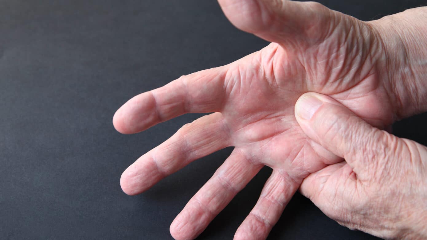
Most people experience a temporary “pins and needles” sensation at some point, such as after sitting in one position for too long or sleeping on an arm. These brief episodes usually resolve once pressure on the affected nerve is relieved.

However, recurring or persistent paresthesia may indicate an underlying condition affecting the nerves, spine, circulation, or musculoskeletal system. This guide explains common causes, associated conditions, treatment options, and how our Relatyv Neurofunctional Pain Management approach may complement your overall treatment plan.

What Is Paresthesia?

Paresthesia is the medical term for abnormal sensations such as tingling, numbness, burning, or “pins and needles.” These sensations most commonly occur in the hands, feet, arms, or legs but may affect other parts of the body.

Temporary paresthesia is often caused by pressure on a nerve that interrupts normal nerve signaling. When symptoms become chronic, frequent, or progressively worsen, they should be evaluated by a healthcare provider.

Common Symptoms

Paresthesia may occur alongside other symptoms, including:

  • Tingling or prickling sensations.
  • Numbness.
  • Burning sensations.
  • Itching.
  • A sensation of the skin “crawling.”
  • Aching or nerve-related pain.
  • Reduced sensation or altered sensitivity to touch.

Conditions Associated with Paresthesia

Peripheral Neuropathy

Peripheral neuropathy occurs when nerves outside the brain and spinal cord become damaged. Common causes include diabetes, vitamin deficiencies, certain medications, alcohol use, and other medical conditions. Symptoms often begin in the feet or hands before gradually progressing.

Nerve Compression

Compressed or pinched nerves may result from herniated discs, spinal stenosis, bone spurs, carpal tunnel syndrome, injuries, poor posture, or repetitive movements.

Poor Circulation

Reduced blood flow caused by vascular disease, diabetes, blood clots, smoking, or prolonged pressure on a limb may contribute to numbness and tingling.

Multiple Sclerosis (MS)

Multiple sclerosis is a neurological condition that damages the protective covering surrounding nerve fibers. Tingling, numbness, and altered sensation are common early symptoms.

Fibromyalgia

Fibromyalgia may cause widespread pain along with tingling, numbness, fatigue, sleep disturbances, and cognitive symptoms.

Sciatica

Sciatica occurs when the sciatic nerve becomes irritated or compressed. Symptoms often include pain, tingling, numbness, or burning sensations that travel from the lower back into the buttock, leg, or foot.

Raynaud’s Phenomenon

Raynaud’s phenomenon causes temporary narrowing of blood vessels, usually in response to cold temperatures or stress, leading to numbness, tingling, color changes, and discomfort in the fingers or toes.

Why Do Damaged Nerves Cause Pins and Needles?

The nervous system carries sensory information between the body and the brain. When nerves become compressed, irritated, inflamed, or damaged, normal communication is disrupted, producing abnormal sensations such as tingling, numbness, burning, or altered sensitivity.

Depending on the underlying condition, symptoms may remain localized or gradually spread to additional areas over time.

When Should You Seek Medical Care?

You should seek medical evaluation if your symptoms:

  • Occur frequently or become progressively worse.
  • Last for weeks or months.
  • Interfere with daily activities.
  • Are accompanied by weakness or reduced range of motion.
  • Occur with persistent pain.
  • Are associated with balance problems or difficulty walking.
  • Are accompanied by changes in bladder or bowel function.

How Paresthesia Is Diagnosed

Your healthcare provider may recommend:

  • Medical history review.
  • Physical and neurological examination.
  • MRI or CT imaging when nerve compression is suspected.
  • Electromyography (EMG) and nerve conduction studies.
  • Blood tests to evaluate for diabetes, vitamin deficiencies, autoimmune disorders, or other medical conditions.
  • Additional testing based on your symptoms and diagnosis.

Conventional Treatment Options

Treatment depends on the underlying cause of your symptoms.

Physical Therapy

Physical therapy may improve strength, flexibility, posture, and movement patterns while reducing pressure on irritated nerves.

Medications

Depending on the diagnosis, medications such as anti-inflammatory drugs, pain relievers, topical medications, or treatments for underlying medical conditions may be recommended. Your healthcare provider can discuss the potential benefits and risks of these options.

Surgery

When significant nerve compression or structural abnormalities are present, surgery may be considered if conservative treatments are unsuccessful.

Our Relatyv Neurofunctional Pain Management Approach

At Relatyv, our Neurofunctional Pain Management approach combines drug-free, non-invasive therapies designed to help reduce chronic pain, improve function, and support healthy nervous system activity.

Each treatment plan is individualized based on your diagnosis, symptoms, and goals. Care may include electroanalgesia, IV therapy when clinically appropriate, nutritional support, and lifestyle recommendations that complement your existing medical care.

While treatment may help reduce pain and improve function for some individuals, outcomes vary depending on the underlying cause of paresthesia.

Address Persistent Tingling and Numbness Early

Recurring numbness, tingling, or burning sensations should not be ignored. Early evaluation can help identify underlying neurological, musculoskeletal, circulatory, or metabolic conditions and guide the most appropriate treatment plan.

At Relatyv, we provide personalized, non-invasive care designed to help reduce chronic pain and improve function. Our Neurofunctional Pain Management approach may complement your existing medical care while supporting your overall mobility and quality of life.

Relatyv - Difficulty Walking

Understanding Factors Behind The Difficulty In Walking

Understanding Factors Behind The Difficulty In Walking 2560 1440 Relatyv

Understanding Difficulty Walking

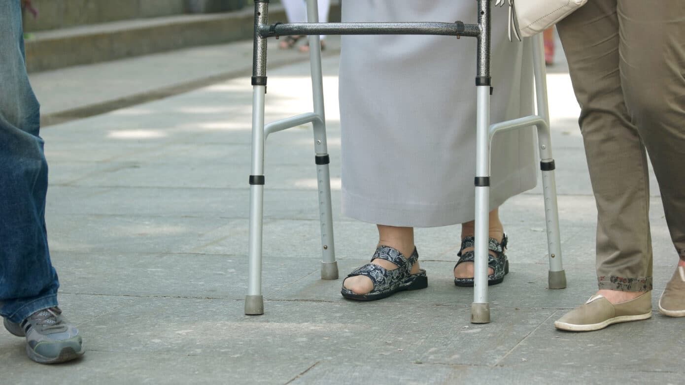
Difficulty walking can affect people of all ages and may develop gradually or appear suddenly following an injury or illness. While temporary mobility problems often improve as the underlying issue heals, persistent changes in your walking pattern may indicate a musculoskeletal, neurological, or circulatory condition that requires medical evaluation.

This guide explains common symptoms, possible causes, treatment options, and how our Relatyv Neurofunctional Pain Management approach may complement your overall treatment plan.

What Does Difficulty Walking Mean?

Difficulty walking, also called a gait abnormality, refers to changes in the way a person walks. These changes may result from pain, weakness, joint instability, nerve dysfunction, balance problems, or limited range of motion.

Depending on the underlying cause, walking difficulties may affect one or both legs and can interfere with daily activities such as climbing stairs, exercising, or standing for prolonged periods.

Common Signs and Symptoms

Walking difficulties may be accompanied by:

  • Limping or an uneven gait.
  • Difficulty maintaining balance.
  • Muscle weakness.
  • Numbness or tingling in the legs or feet.
  • Hip, knee, ankle, or foot pain.
  • Lower back pain.
  • Leg cramps or muscle spasms.
  • Joint stiffness.
  • Reduced endurance when walking.

Common Types of Gait Abnormalities

Healthcare providers may describe abnormal walking patterns using specific terms, including:

  • Steppage gait: Difficulty lifting the front of the foot while walking.
  • Spastic gait: Stiff, dragging movements of the legs.
  • Waddling gait: Side-to-side shifting of the hips while walking.
  • Propulsive gait: A forward-leaning posture with short, rapid steps.
  • Scissor gait: Legs crossing over one another during walking.

Conditions That May Cause Difficulty Walking

Injuries

Fractures, sprains, strains, ligament tears, tendon injuries, and joint dislocations can temporarily limit mobility until healing occurs.

Spinal Stenosis

Narrowing of the spinal canal may compress nerves traveling to the legs, leading to pain, weakness, numbness, or difficulty walking.

Joint Instability

Damaged ligaments, tendon injuries, muscle weakness, or cartilage loss may create a feeling that the knee, ankle, or hip is unstable during movement.

Gout

Inflammatory arthritis caused by uric acid crystal deposits commonly affects the feet, ankles, and knees, making walking painful during flare-ups.

Foot and Ankle Conditions

Conditions such as plantar fasciitis, Achilles tendinopathy, flat feet, heel spurs, and other structural foot abnormalities may alter walking mechanics.

Osteoarthritis

Cartilage loss within weight-bearing joints may cause pain, stiffness, swelling, and reduced mobility that affects walking.

Rheumatoid Arthritis

Inflammation associated with rheumatoid arthritis frequently affects the knees, ankles, and feet, making standing and walking more difficult.

Neurological Disorders

Conditions affecting the nervous system—including peripheral neuropathy, multiple sclerosis, Parkinson’s disease, spinal cord injuries, and stroke—may impair balance, coordination, muscle strength, and sensation.

Peripheral Artery Disease (PAD)

Reduced blood flow to the legs may cause cramping, weakness, fatigue, and pain during walking.

Lifestyle Factors That May Affect Mobility

Several modifiable factors can contribute to walking difficulties, including:

  • Physical inactivity.
  • Excess body weight.
  • Muscle weakness.
  • Poor balance.
  • Smoking.
  • Excessive alcohol use.
  • Poor nutrition.
  • Delayed rehabilitation following injury or surgery.

When Should You Seek Medical Care?

You should seek medical evaluation if walking difficulties:

  • Continue to worsen.
  • Interfere with daily activities.
  • Are accompanied by persistent pain.
  • Occur with numbness or weakness.
  • Cause repeated falls or balance problems.
  • Develop after an injury.

Seek emergency medical care immediately if walking difficulties develop suddenly along with:

  • Facial drooping.
  • Arm or leg weakness.
  • Difficulty speaking.
  • Sudden vision changes.
  • Confusion.
  • A severe, sudden headache.

These symptoms may indicate a stroke or another medical emergency.

How Difficulty Walking Is Diagnosed

Your healthcare provider may recommend:

  • Medical history review.
  • Physical examination.
  • Neurological assessment.
  • Gait analysis.
  • X-rays, MRI, or CT imaging.
  • Blood tests when inflammatory or metabolic conditions are suspected.
  • Nerve conduction studies or electromyography (EMG) when nerve disorders are suspected.

Conventional Treatment Options

Treatment depends on the underlying diagnosis.

Medications

Medications may be prescribed to manage pain, reduce inflammation, or treat underlying medical conditions. Your healthcare provider can discuss the potential benefits and risks based on your specific diagnosis.

Physical Therapy

Physical therapy can improve strength, flexibility, balance, coordination, and walking mechanics while supporting recovery after injury or illness.

Assistive Devices

Canes, walkers, braces, or other mobility aids may improve safety and independence when appropriate.

Lifestyle Modifications

Regular exercise, weight management, smoking cessation, balanced nutrition, and ongoing rehabilitation may improve mobility and reduce strain on the joints.

Surgery

In some cases, surgery may be recommended to repair damaged joints, relieve nerve compression, or correct structural problems when conservative treatment has not been successful.

Our Relatyv Neurofunctional Pain Management Approach

At Relatyv, our Neurofunctional Pain Management approach combines drug-free, non-invasive therapies designed to help reduce chronic pain, improve function, and support healthy nervous system activity.

Each treatment plan is individualized based on your diagnosis, symptoms, and goals. Care may include electroanalgesia, IV therapy when clinically appropriate, nutritional support, and lifestyle recommendations that complement your existing medical care.

Improve Mobility with Individualized Care

Difficulty walking can result from many different medical conditions, making an accurate diagnosis an important first step. Early evaluation may help identify treatable causes and reduce the risk of worsening mobility or falls.

At Relatyv, we provide personalized, non-invasive care designed to help reduce chronic pain and improve function. Our Neurofunctional Pain Management approach may complement your existing treatment plan while supporting greater mobility and overall quality of life.

Relatyv - Pupil Constriction

Pupil Constriction Induced By Cluster Headaches

Pupil Constriction Induced By Cluster Headaches 10000 5633 Relatyv

Cluster Headaches and Pupil Constriction (Miosis)

Cluster headaches are a rare but severe type of headache characterized by intense pain on one side of the head, often occurring in recurring cycles or “clusters.” During an attack, some people also experience changes involving the eye on the affected side, including pupil constriction (miosis), tearing, redness, or a drooping eyelid.

This guide explains why pupil constriction occurs with cluster headaches, associated symptoms, treatment options, and how our Relatyv Neurofunctional Pain Management approach may complement your overall treatment plan.

What Is Pupil Constriction?

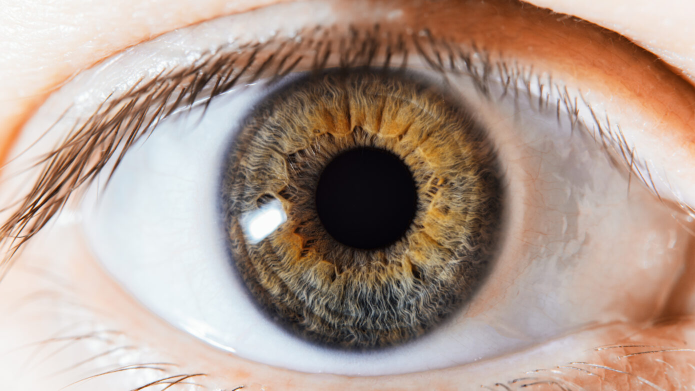
Pupil constriction, also known as miosis, occurs when the opening in the center of the eye becomes smaller. Normally, the pupils continuously adjust to changes in lighting conditions.

During a cluster headache attack, changes affecting the autonomic nervous system and nearby nerves may cause one pupil to remain smaller than usual for the duration of the attack.

Why Does Miosis Occur During Cluster Headaches?

Several factors may contribute to pupil constriction during a cluster headache, including:

  • Activation of the autonomic nervous system.
  • Involvement of the trigeminal nerve.
  • Changes affecting nearby blood vessels and surrounding tissues.

These neurological changes can contribute to both the severe headache pain and the eye-related symptoms experienced during an attack.

Symptoms Associated with Pupil Constriction

Along with severe one-sided head pain, people experiencing miosis during a cluster headache may notice:

  • A smaller pupil on the affected side.
  • Redness of the eye.
  • Excessive tearing.
  • A drooping eyelid (ptosis).
  • Blurred or double vision.
  • Difficulty focusing.
  • Sensitivity to light.

How Pupil Constriction Can Affect Daily Life

Although headache pain is typically the most disabling symptom, changes in vision can also interfere with everyday activities. Reading, driving, working, or using digital devices may become more difficult during or shortly after a cluster headache attack.

Recurring headaches can also affect sleep, work performance, and overall quality of life.

When Should You See a Healthcare Provider?

You should seek medical evaluation if you experience:

  • New or recurring episodes of one-sided pupil constriction.
  • Persistent differences in pupil size.
  • Pupil changes accompanied by severe headaches.
  • Drooping eyelid, excessive tearing, or redness of the eye.
  • Vision changes during headache episodes.

Seek emergency medical care immediately if sudden pupil changes occur with weakness, confusion, loss of consciousness, difficulty speaking, or sudden vision loss, as these symptoms may indicate a medical emergency.

How Cluster Headaches Are Diagnosed

Your healthcare provider may recommend:

  • Medical history review.
  • Physical and neurological examination.
  • Detailed review of headache patterns and associated symptoms.
  • MRI or CT imaging when necessary to rule out other neurological conditions.

Because several headache disorders can produce similar symptoms, obtaining an accurate diagnosis is an important first step in developing an appropriate treatment plan.

Conventional Treatment Options

Treatment focuses on relieving attacks and reducing the frequency of future episodes.

Acute Treatments

Depending on your individual needs, your healthcare provider may recommend treatments such as high-flow oxygen therapy or medications that help shorten a cluster headache attack.

Preventive Medications

Medications may be prescribed to reduce how often cluster headaches occur. Your healthcare provider can discuss which options may be appropriate based on your medical history.

Complementary Therapies

Some individuals may also incorporate therapies such as acupuncture, stress management, or other supportive approaches as part of a comprehensive care plan. Discuss these options with your healthcare provider before beginning treatment.

Our Relatyv Neurofunctional Pain Management Approach

At Relatyv, our Neurofunctional Pain Management approach combines drug-free, non-invasive therapies designed to help reduce chronic pain, improve function, and support healthy nervous system activity.

Each treatment plan is individualized based on your diagnosis, symptoms, and goals. Care may include electroanalgesia, IV therapy when clinically appropriate, nutritional support, and lifestyle recommendations that complement your existing medical care.

While treatment may help reduce headache-related pain and improve function for some individuals, outcomes vary depending on the underlying condition and individual response to care.

Managing Cluster Headaches with Individualized Care

Cluster headaches and associated eye symptoms can significantly affect daily life, but effective management begins with an accurate diagnosis and a personalized treatment strategy.

At Relatyv, we provide personalized, non-invasive care designed to help reduce chronic pain and improve function. Our Neurofunctional Pain Management approach may complement your existing treatment plan while supporting your overall health and quality of life.

Relatyv - Digestive Pain

Digestive Issues

Digestive Issues 2560 1442 Relatyv

Understanding Digestive Issues

Digestive issues are common and can affect people of all ages. Occasional symptoms such as constipation, diarrhea, bloating, or nausea are often temporary, but persistent digestive problems may indicate an underlying medical condition that should be evaluated by a healthcare provider.

This guide explains common digestive symptoms, possible causes, treatment options, and how our Relatyv Neurofunctional Pain Management approach may complement your overall treatment plan.

Common Digestive Symptoms

Digestive problems can vary widely depending on the underlying cause. Common symptoms include:

Constipation

Constipation involves infrequent, difficult, or painful bowel movements. It may result from dehydration, dietary changes, medications, hormonal changes, or certain medical conditions.

Diarrhea

Diarrhea is characterized by frequent loose or watery stools. It may be caused by infections, food intolerances, medications, stress, or chronic gastrointestinal conditions.

Abdominal Pain and Nausea

Abdominal discomfort and nausea can occur for many reasons, including digestive disorders, infections, food sensitivities, inflammation, or other medical conditions.

Irregular Bowel Movements

Changes in bowel habits—including stool frequency, consistency, or urgency—may be temporary or may signal an underlying digestive disorder when persistent.

Gas and Bloating

Gas and bloating commonly occur after eating certain foods or large meals, but ongoing symptoms without an obvious cause should be discussed with your healthcare provider.

Conditions That May Cause Digestive Symptoms

Inflammatory Bowel Disease (IBD)

Inflammatory bowel disease is a chronic condition involving inflammation of the digestive tract. The two primary forms are Crohn’s disease and ulcerative colitis.

Ulcerative Colitis

Ulcerative colitis affects the lining of the large intestine and may cause abdominal pain, diarrhea, urgency, rectal bleeding, and fatigue.

Crohn’s Disease

Crohn’s disease can affect any portion of the digestive tract and may cause abdominal pain, diarrhea, weight loss, bloating, nausea, and nutritional deficiencies.

Other Chronic Conditions

Digestive symptoms may also occur alongside other chronic medical conditions. For example, some individuals with endometriosis experience abdominal pain, bloating, constipation, diarrhea, or nausea related to their condition.

Home Strategies That May Help

Depending on the cause of your symptoms, healthy lifestyle habits may support digestive function, including:

  • Regular physical activity.
  • Eating a balanced diet with appropriate fiber intake.
  • Drinking adequate water throughout the day.
  • Identifying and avoiding foods that trigger symptoms.
  • Managing stress.
  • Gentle abdominal massage when recommended by your healthcare provider.

If symptoms persist or worsen, medical evaluation is recommended.

When Should You See a Healthcare Provider?

Seek medical evaluation if you experience:

  • Blood in your stool.
  • Unexplained weight loss.
  • Persistent diarrhea or constipation.
  • Abdominal pain that wakes you during the night.
  • A family history of inflammatory bowel disease or other gastrointestinal disorders.
  • Digestive symptoms that interfere with your daily life.

How Digestive Problems Are Diagnosed

Your healthcare provider may recommend:

  • Medical history review.
  • Physical examination.
  • Blood tests.
  • Stool testing.
  • Imaging studies when appropriate.
  • Endoscopy or colonoscopy if indicated.
  • Referral to a gastroenterologist for specialized evaluation.

An accurate diagnosis helps determine the most appropriate treatment based on the underlying cause of your symptoms.

Conventional Treatment Options

Treatment depends on the specific digestive condition being treated.

Medications

Your healthcare provider may recommend medications to manage symptoms such as diarrhea, constipation, abdominal cramping, inflammation, or acid-related disorders. The benefits and potential risks of these treatments should be discussed based on your individual diagnosis.

Dietary Changes

Nutrition modifications, including adjusting fiber intake or avoiding foods that worsen symptoms, are often an important part of digestive health management.

Surgery

For certain severe gastrointestinal conditions, surgery may be considered when conservative treatments have not been successful or when complications develop.

Our Relatyv Neurofunctional Pain Management Approach

At Relatyv, our Neurofunctional Pain Management approach combines drug-free, non-invasive therapies designed to help reduce chronic pain, improve function, and support healthy nervous system activity.

Each treatment plan is individualized based on your diagnosis, symptoms, and goals. Care may include electroanalgesia, IV therapy when clinically appropriate, nutritional support, and lifestyle recommendations that complement your existing medical care.

For individuals experiencing chronic pain conditions that may contribute to digestive discomfort, this approach may help reduce pain and improve overall function as part of a comprehensive treatment plan.

Take Ongoing Digestive Symptoms Seriously

While occasional digestive upset is common, persistent or worsening symptoms should not be ignored. Early evaluation can help identify underlying gastrointestinal or systemic conditions and guide the most appropriate treatment.

At Relatyv, we provide personalized, non-invasive care designed to help reduce chronic pain and improve function. Our Neurofunctional Pain Management approach may complement your existing treatment plan while supporting your overall health and quality of life.

Relatyv - Numbness

Experiencing A Sensation Of Numbness In The Body

Experiencing A Sensation Of Numbness In The Body 2560 1442 Relatyv

Understanding Numbness

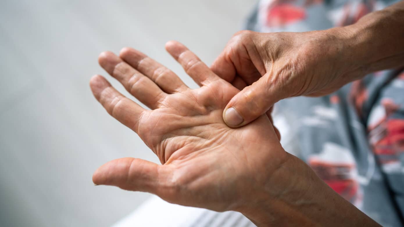
Numbness is a common symptom that many people experience at some point in their lives. Often described as a “pins and needles” sensation or loss of feeling, numbness can affect the hands, feet, arms, legs, face, or other areas of the body.

Temporary numbness may occur after sitting or sleeping in one position for too long. However, persistent, recurring, or worsening numbness may indicate an underlying neurological, musculoskeletal, or circulatory condition that should be evaluated by a healthcare provider.

This guide explains common causes of numbness, associated medical conditions, treatment options, and how our Relatyv Neurofunctional Pain Management approach may complement your overall treatment plan.

What Is Numbness?

Numbness, also called paresthesia, refers to reduced or altered sensation in part of the body. It is often accompanied by tingling, prickling, burning, or a “pins and needles” feeling.

These sensations occur when normal communication between the nerves and the brain is disrupted. Depending on the cause, numbness may be temporary or persistent.

Common Causes of Numbness

Many factors can contribute to numbness, including:

  • Poor circulation.
  • Pinched or compressed nerves.
  • Back or neck problems, such as herniated discs or spinal stenosis.
  • Injuries affecting nerves or surrounding tissues.
  • Poor posture.
  • Repetitive movements that place pressure on nerves.

Medical Conditions Associated with Numbness

Sciatica

Sciatica occurs when the sciatic nerve becomes irritated or compressed, causing pain, tingling, or numbness that travels from the lower back into the leg or foot.

Peripheral Neuropathy

Peripheral neuropathy involves damage to nerves outside the brain and spinal cord and commonly causes numbness, tingling, burning, or weakness in the hands and feet.

Diabetic Neuropathy

Long-term diabetes may damage peripheral nerves, leading to reduced sensation, numbness, and pain, particularly in the feet and lower legs.

Fibromyalgia

Fibromyalgia is a chronic pain condition that may cause widespread pain, fatigue, and intermittent numbness or tingling sensations.

Multiple Sclerosis (MS)

Multiple sclerosis is a neurological disorder that damages nerve fibers and may produce numbness, weakness, balance problems, and changes in coordination.

Tarsal Tunnel Syndrome

Compression of the tibial nerve near the ankle may cause numbness, tingling, or burning sensations in the foot.

Peroneal Nerve Dysfunction

Damage or compression of the peroneal nerve may lead to numbness, weakness, or difficulty lifting the foot.

Peripheral Artery Disease (PAD)

Reduced blood flow to the legs caused by peripheral artery disease may contribute to numbness, pain, and cramping during activity.

Trigeminal Neuralgia

Although trigeminal neuralgia is best known for episodes of severe facial pain, some individuals may also experience altered sensation or numbness in the face.

When Should You Seek Medical Care?

You should seek medical evaluation if numbness:

  • Affects only one side of the body.
  • Occurs with muscle weakness or paralysis.
  • Persists or becomes progressively worse.
  • Is accompanied by severe pain.
  • Interferes with daily activities.

Seek emergency medical care immediately if numbness develops suddenly along with:

  • Difficulty speaking.
  • Confusion.
  • Vision changes.
  • Dizziness.
  • Facial drooping.
  • Sudden weakness.

These symptoms may indicate a stroke or another medical emergency.

How Numbness Is Diagnosed

Your healthcare provider may recommend:

  • Medical history review.
  • Physical and neurological examination.
  • Blood tests.
  • MRI or CT imaging when appropriate.
  • Nerve conduction studies or electromyography (EMG).

An accurate diagnosis helps determine the most appropriate treatment for your symptoms.

Conventional Treatment Options

Treatment depends on the underlying cause of the numbness.

Medications

Your healthcare provider may recommend medications to help manage pain, inflammation, or underlying medical conditions. The benefits and potential risks of these treatments should be discussed based on your individual diagnosis.

Physical Therapy

Physical therapy may improve strength, flexibility, posture, and movement while reducing pressure on affected nerves and supporting recovery.

Injections

For some conditions involving inflammation or nerve compression, corticosteroid injections may be recommended as part of an overall treatment plan.

Surgery

When significant nerve compression or structural abnormalities are present, surgery may be considered if conservative treatments are unsuccessful.

Our Relatyv Neurofunctional Pain Management Approach

At Relatyv, our Neurofunctional Pain Management approach combines drug-free, non-invasive therapies designed to help reduce chronic pain, improve function, and support healthy nervous system activity.

Each treatment plan is individualized based on your diagnosis, symptoms, and goals. Care may include electroanalgesia, IV therapy when clinically appropriate, nutritional support, and lifestyle recommendations that complement your existing medical care.

While treatment may help reduce pain and improve function for some individuals, outcomes vary depending on the underlying cause of numbness.

Address Persistent Numbness Early

Persistent or recurring numbness should not be ignored. Early evaluation can help identify neurological, musculoskeletal, circulatory, or metabolic conditions that may benefit from treatment.

At Relatyv, we provide personalized, non-invasive care designed to help reduce chronic pain and improve function. Our Neurofunctional Pain Management approach may complement your existing treatment plan while supporting your overall health and quality of life.

Relatyv - Range of Motion

Limited Range of Motion in Knee: What Does It Mean?

Limited Range of Motion in Knee: What Does It Mean? 2560 1442 Relatyv

Understanding Limited Knee Range of Motion

Your knees allow you to walk, climb stairs, squat, and perform countless everyday activities. When your knee cannot fully bend or straighten, these movements become more difficult and may affect your independence, mobility, and quality of life.

While temporary stiffness after an injury is common, persistent loss of knee range of motion may indicate an underlying musculoskeletal or neurological condition that should be evaluated by a healthcare provider.

This guide explains common causes of limited knee mobility, treatment options, and how our Relatyv Neurofunctional Pain Management approach may complement your overall treatment plan.

What Is Limited Knee Range of Motion?

Range of motion (ROM) refers to how far your knee can bend (flexion), straighten (extension), and rotate during movement. Limited range of motion means the knee cannot move through its normal range due to pain, stiffness, swelling, weakness, injury, or joint changes.

Loss of mobility may be temporary or long-lasting, depending on the underlying cause.

Symptoms of Limited Knee Mobility

People with reduced knee range of motion may experience:

  • Difficulty fully bending or straightening the knee.
  • Stiffness.
  • Pain during movement.
  • Swelling.
  • Clicking or catching sensations.
  • Weakness.
  • Difficulty walking, climbing stairs, or standing from a seated position.

Common Causes of Limited Knee Range of Motion

Chronic Knee Pain

Long-term knee pain may develop from overuse, injuries, arthritis, or other musculoskeletal conditions. Pain and inflammation often make movement uncomfortable, contributing to stiffness and reduced mobility.

Osteoarthritis

Osteoarthritis gradually wears away cartilage within the knee joint, leading to pain, stiffness, swelling, and decreased flexibility.

Injuries

Fractures, ligament tears, tendon injuries, cartilage damage, and dislocations may temporarily or permanently affect knee movement, particularly if rehabilitation is delayed.

Infections

Joint infections, including septic arthritis, can cause pain, swelling, warmth, and difficulty moving the knee. These conditions require prompt medical evaluation.

Muscle Weakness or Tightness

Weak or tight muscles surrounding the knee may limit flexibility and reduce the joint’s ability to move normally.

Neurological Conditions

Conditions such as multiple sclerosis, stroke, spinal cord injuries, cerebral palsy, and certain neuromuscular disorders may affect muscle control and limit knee movement.

When Should You Seek Medical Care?

You should seek medical evaluation if you:

  • Cannot fully bend or straighten your knee.
  • Experience recurring stiffness or loss of motion.
  • Have pain or swelling that does not improve.
  • Find daily activities becoming increasingly difficult.
  • Develop redness, warmth, or fever along with knee pain.

Prompt evaluation is especially important if symptoms follow a significant injury or develop suddenly.

How Limited Knee Range of Motion Is Diagnosed

Your healthcare provider may recommend:

  • Medical history review.
  • Physical examination.
  • Measurement of knee range of motion.
  • X-rays or other imaging studies.
  • MRI when soft tissue injuries are suspected.
  • Additional testing based on your symptoms and diagnosis.

Conventional Treatment Options

Treatment depends on the underlying cause of the limited mobility.

Rest and Home Care

For minor injuries, rest, ice, compression, elevation, and activity modification may reduce swelling and improve mobility during recovery.

Physical Therapy

Physical therapy may improve flexibility, strength, balance, and knee function while supporting recovery after injury or surgery.

Shockwave Therapy

Extracorporeal shockwave therapy (ESWT) may be recommended for certain musculoskeletal conditions as part of a broader treatment plan when appropriate.

Medications

Your healthcare provider may recommend medications to help manage pain or inflammation while treating the underlying condition. The potential benefits and risks vary depending on your diagnosis.

Surgery

For severe joint damage or structural abnormalities, surgical procedures such as ligament reconstruction or knee replacement may be considered when conservative treatments have not provided adequate improvement.

Our Relatyv Neurofunctional Pain Management Approach

At Relatyv, our Neurofunctional Pain Management approach combines drug-free, non-invasive therapies designed to help reduce chronic pain, improve function, and support healthy nervous system activity.

Each treatment plan is individualized based on your diagnosis, symptoms, and goals. Care may include electroanalgesia, IV therapy when clinically appropriate, nutritional support, and lifestyle recommendations that complement your existing medical care.

While treatment may help reduce pain and improve function for some individuals, outcomes vary depending on the underlying cause of reduced knee mobility.

Improve Knee Mobility with Individualized Care

Limited knee range of motion can make everyday activities more challenging, but early evaluation can help identify the cause and guide appropriate treatment.

At Relatyv, we provide personalized, non-invasive care designed to help reduce chronic pain and improve function. Our Neurofunctional Pain Management approach may complement your existing treatment plan while supporting greater mobility and overall quality of life.

Relatyv - Range of Motion

Limited Range of Motion in Knee: What Does It Mean?

Limited Range of Motion in Knee: What Does It Mean? 2560 1440 Relatyv

In our everyday life, we perform movements such as walking, jogging, climbing stairs, or bending down to tie our shoes almost automatically. And, if we are able to perform these movements, it is thanks to the knee’s complex structure of bones, ligaments, tendons, cartilage, and muscles, which gives it its exceptional mobility.

But it is often too easy to take such a range of motion for granted. Over time, injuries, diseases, and health conditions can significantly reduce your knees’ range of motion and have a profound impact on your ability to remain functional in your daily life.

Fortunately, undergoing extensive knee surgeries and enduring the consequences of invasive procedures are no longer the only options to restore your range of motion and knee health.

With the whole-person approach pioneered–Neuragenex Neurofunctional Pain Management – you can regain your knee health without surgical interventions or medications. Let’s discover how in this guide.

What Happens When The Range Of Motion In The Knee Is Reduced?

The knee plays a vital role in bearing and redistributing loads during walking or running, absorbing shock, and balancing our body as it moves through space. This complex joint links the upper leg bone (the femur or thigh bone) and the lower leg bones (fibula and tibia, or shin bone), which are protected by a frontal flat bone called the patella

These bones are connected by four ligaments:

  • Anterior cruciate ligament (ACL)
  • Posterior cruciate ligament (PCL)
  • Medial collateral ligament (MCL)
  • Lateral collateral ligament (LCL)

Each bone is also connected to the muscles of the upper and lower leg by tendons. The knee joint is kept lubricated and protected from friction damage by cartilaginous tissues, which act as shock-absorbing “cushions”.

This complex structure enables the three main movements of the knee:

  • Extension (straightening the knee)
  • Flexion (bending the knee)
  • Lateral rotation

Full range of motion (ROM) of the knee refers to its full movement potential. Limited ROM refers to a condition where the knee joint is unable to move through its full range of motion.

Limited ROM may occur due to infection, injury, mechanical issues, inflammation, and degenerative diseases like osteoarthritis. According to estimations, as much as a quarter of the population may suffer from a reduced range of motion to some extent.

A slight or short-term reduction in knee ROM may be due to poor muscle conditioning or minor injuries. These are temporary conditions that can be addressed easily or resolved by themselves with at-home care.

However, in some cases, the inability to move the knee through its full range of motion is a symptom of an underlying condition – such as arthritis – that requires immediate medical attention.

When left unaddressed, limited ROM can have devastating consequences, including:

  • Limiting your ability to remain functional and active in your daily life
  • Pain, stiffness, and further loss of range of motion
  • Significant impairment and disability
  • Preventing you from living independently or aging in place
  • If present in only one knee, it can lead to imbalances, mechanical issues, and poor posture
  • May lead to relative disuse of one knee, and subsequent loss of muscle strength

For athletes and sportspeople, limited range of motion may also impact the outcome of resistance training, leading to decreased performance.

Conditions Associated With Limited Knee Mobility

As we get older, the components of the knee may suffer progressive degeneration, which translates into a normal, age-related loss of range of motion. However, some conditions can impact your knee health and reduce your knee mobility drastically.

Below, we’ll look at the health events and diseases that may compromise your knee’s ability to move as it should.

Chronic Knee Pain

Chronic knee pain is a condition that often arises from degenerative conditions, traumatic injuries, overuse, or mechanical issues. If left unaddressed, chronic knee pain can lead to knee disuse, muscle atrophy, and disability.

It is estimated that one in four adults suffer from long-term knee pain, and this prevalence is on the rise due to the aging of the population and increasing rates of obesity.

Chronic knee pain is often characterized by an excessive and sustained inflammatory response. In turn, high levels of inflammation can lead to swelling, pain, and stiffness, which can restrict joint movement and prevent the knee from flexing or extending to its full range of motion.

Osteoarthritis

Osteoarthritis (OA) is an inflammatory condition of the joints that lead to the progressive degeneration of the cartilage. As the cartilage breaks down, the joint’s components are exposed to friction and shock damage during movement, which leads to damage to bones, ligaments, and tendons.

Over time, osteoarthritis can cause the formation of abnormal bone growths (bone spurs) and deformities, which prevent the knee from moving as it normally should.

Often caused by natural age-related wear-and-tear, overuse, and poor mechanics, OA is the most common form of arthritis and the leading cause of disability in the elderly. That’s because, if left unaddressed, osteoarthritis can compromise the mechanics of the knee joint and cause irreversible damage to the components responsible for safeguarding its full range of motion.

Like other forms of arthritis, OA also causes joint pain, stiffness, swelling, clicking noises, and “catching” sensations, which can make knee movements even more undesirable.

Injuries And Tissue Damage

Traumatic injuries that cause damage to tissues like bones, ligaments, and tendons can have a long-lasting impact on the knee’s ROM. This can occur for multiple reasons:

  • An acute knee injury can permanently compromise one or more of the components of the knees
  • Lack of rehabilitation after an injury or surgical procedure can cause long-lasting damage
  • Injury and immobilization can lead to relative disuse of the knee, which translates into a subsequent loss of muscle mass and strength
  • Abnormal scar tissue can build up after a fracture or surgical intervention (i.e.: after an anterior cruciate ligament reconstruction), binding components of the knee together and limiting their mobility. This condition is known as arthrofibrosis.
  • Unsuccessful surgical interventions or inadequate recovery from a broken bone can impact normal knee mechanics and expose the joint components to excessive stress.

Some injuries that can lead to a loss of ROM include dislocations and fractures, as well as torn ligaments, tendons, and cartilage.

Infections

Some infections caused by bacteria, viruses, and fungi tend to target the joint’s structures. For example, in children, hip infections can lead to pain, swelling, and subsequent range of motion.

In the knee, the most common infection is septic or viral arthritis. This condition occurs when germs and viruses travel through the bloodstream from other infected areas of the body (i.e.: following an animal bite or penetrating injury). Some of the viruses that may cause septic arthritis include hepatitis B, hepatitis C, and the Zika virus.

Septic arthritis causes symptoms like swelling, pain, and stiffness, which translates into difficulty moving the joint and a reduced range of motion. Around 20,000 cases of septic arthritis are reported in the US alone each year.

Muscle Weakness

The muscles of the upper and lower leg play a vital role in initiating and controlling knee movements. When these muscles lose mass, strength, or flexibility, you may struggle to bend or extend the knee fully.

Some conditions associated with reduced ROM include:

  • Muscle tightness. Thigh muscles can impact the flexibility of the knee, preventing it from stretching or flexing as it should.
  • Muscle weakness. Without enough muscle power, you may be unable to extend or bend your knee, as well as keep it bent or extended.
  • Muscle atrophy. A severe loss of muscle mass that occurs after periods of immobilization or disuse can significantly compromise knee mobility.

Poor muscle conditioning can create a vicious cycle that can further restrict your ROM: weak muscles may not support daily movements like climbing stairs, walking, or jogging, which, in turn, can lead to a loss of strength and mobility.

Neurological/Neuromuscular Conditions

Some neurological and neuromuscular conditions interfere with the normal transmission and process of nerve signals relating to muscle movements. When these signals are unable to reach the brain or are not properly processed, you may become unable to control muscle movements.

Disorders that cause damage to muscles, the brain, or the nerves in the central and peripheral nervous system include:

  • Cerebral palsy
  • Spinal cord injury
  • Multiple sclerosis
  • Stroke
  • Brain or head injury
  • Muscle dystrophy
  • Contracture deformities (conditions that cause components of the joint to become permanently fixed)

Without adequate care, these conditions can lead to severe complications, including muscle atrophy, numbness, and disability.

When Should I See My Doctor?

If you have experienced a minor trauma – such as a knee ligament sprain – your loss of range of movement will be a temporary condition, mostly associated with the inflammatory response of the body to injury. In this case, at-home care will be enough to ease pain and swelling and help you regain your knee function in 2-4 weeks.

However, if you suffer from a degenerative joint condition or chronic pain, the loss of range of movement can be recurring, long-lasting, and incredibly impactful on your day-to-day life. What’s more, reduced knee mobility may be a symptom of an underlying neurological disease, which requires immediate medical attention.

Generally, you should see your doctor if:

  • You can’t fully straighten or bend your knees
  • You frequently suffer from reduced knee mobility or recurring ROM problems
  • Your daily activities are affected by the loss of ROM

It is important to note that, as you age, you are likely to lose joint mobility to some extent.

Although this is normal, you should be able to retain what’s known as a “functional range of movement”. This is the minimum range of movement your knee needs to allow for everyday activities, like climbing up stairs, bending, walking, sitting down, standing up from sitting, riding a bike, squatting, and tying your shoelaces.

A healthy knee’s full range of motion is 135 degrees. Most daily activities require full extension but only 110-120 degrees of knee flexion. In turn, losing a few degrees of knee flexion won’t affect your daily activities, while not being able to fully extend your knee can significantly impact your mobility.

Measuring And Diagnosing Limited Range Of Motion

To understand how a limited ROM is diagnosed and measured, it is firstly important to clarify the difference between active, passive, and active assisted range of motion. Let’s look at these different ways of measuring ROM below:

  • Active ROM: Active range of motion refers to how much you can flex and extend the knee autonomously. When assessing active ROM, you’ll be bending and extending your knee by contracting your muscles, without external help.
  • Passive ROM: Passive ROM is the measure of how much your knee can flex and extend when someone moves or stretches your leg. A physician or specialized machine will help you bend and extend your knee if you are unable to do so by contracting your muscles.
  • Active assisted ROM: Active assisted ROM measures how much you can move your knee with some assistance.

Although it is normal for passive knee ROM to be greater than active ROM, these two measures should be similar in a healthy, pain-free knee.

According to a study conducted by the CDC on the range of motion of different joints, a healthy knee should be able to move as follow:

  Age 20–44 Age 45–69
Motion Females Males Females Males
Knee Flexion 141.9° 137.7° 137.8° 132.9°
Knee Extension 1.6° 1.0° 1.2 ° 0.5°

The knee should also be able to rotate internally by 10 degrees and externally by 30-40 degrees. Normal passive knee flexion can be as much as 150°, while passive knee extension can be up to 10° of hyperextension.

Given these values, your doctor will determine the extent of your loss of ROM and diagnose any underlying condition that may be aggravating your knee mobility

Some of the diagnostic tools and techniques used in the process include:

  • Medical exam: Your doctor will carry out a physical examination, a review of your symptoms, and assessments of your medical history.
  • Imaging tests: Imaging tests like X-rays may be used to obtain a clear picture of the inside of your knee, and to determine the presence of abnormal tissue (i.e.: tumors) and mechanical issues.
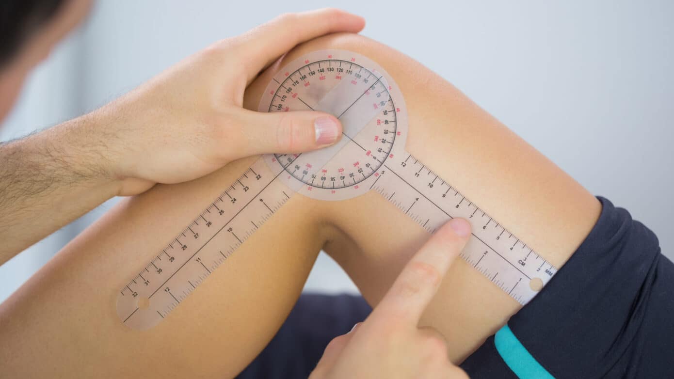
  • Goniometry assessment: Using a goniometer is the most accurate way to diagnose the extent of range of motion loss. A goniometer is an instrument designed for the measurements of angles. It is composed of a central round body representing 360 degrees of motion and two arms, one stationary and one moveable.

To measure your range of knee flexion and extension, your doctor will carry out the following steps:

  1. He or she will ask you to lie on your back on a flat surface and straighten your leg
  2. The goniometer will be positioned so that the central body of the instrument is against the side of the knee, on the lateral femoral epicondyle (the outer part of the knee).
  3. Keeping the goniometer in place, your physician will set the stationary arm along the outer thigh, in line with the greater trochanter (the bony lump located at the top of the thigh)
  4. The moveable arm of the goniometer will be set along the lower leg, in line with the lateral malleolus of the ankle (the bony lump visible on the outer part of the ankle).
  5. The physician will ask you to use your muscles to straighten the leg and measure the knee extension
  6. To measure knee flexion, your doctor will ask you to bend your knee as far as you can by bringing your foot closer to the buttocks.

  • Self-assessment: You can get an estimation of your knee flexion and extension without a goniometer. To do so follow these steps:
  1. Lie on your back on a flat and firm surface
  2. Straighten the leg using your muscles and try to slide your hand underneath the knee
  3. If you can’t fit any of your fingers, your knee is hyperextended. If you can slide all of your fingers under the knee, you are lacking ≈5 degrees in extension
  4. To estimate knee flexion, slide your foot along the surface towards your buttocks. Then, measure the distance between your buttocks and your heel. This method can be used to check your progress and try to improve knee flexion, but won’t give you a measurement.
  • Other methods: To obtain a more accurate measurement of your knee range of motion at home, you can download a goniometer app. You may ask a friend or family member to help you measure your knee extension and flexion.


Conventional Treatments For Limited Knee Range Of Motion


As seen above, treating a temporary reduction in range of motion is a matter of finding the right at-home remedies, such as rest, icing, and compression. However, if your mobility issue is long-lasting or recurring, your first port of call should be obtaining an accurate diagnosis.

Once you and your physician have an understanding of what’s affecting your knee, you may be recommended one or more of the following treatments:

  • Compresses: Icing a new injury such as a sprain can reduce swelling and stiffness by preventing the build-up of fluids around the injured tissues.

In the case of old injuries that still cause pain, chronic pain conditions, or inflammatory diseases like arthritis, hot compresses can support the circulation of blood and nutrients and relax tense muscles.

Ultimately, compresses can ease down painful flare-ups, which may otherwise get in the way of your knee mobility.

  • Exercise: If the loss of ROM is caused by muscle weakness or tightness, you may benefit from physical therapy and exercise. A therapist will guide you in the process of exercising your knee, avoiding further injuries, improving flexibility, and rehabilitating weak muscles.
  • Shockwave Therapy: Extracorporeal Shockwave Therapy (ESWT) is a form of treatment that uses acoustic waves delivered through the skin. The low-frequency waves support the healing of damaged tissues, promote circulation, and stimulate nerves at the site of injury.

According to a 2020 study, this therapy can help improve ROM in people with knee osteoarthritis.

  • Surgery: Diseases such as arthritis and traumatic injuries may severely compromise the components of the knee. Surgical interventions like knee arthroplasty may be used to replace these worn-out components with an artificial joint.

Given the lengthy rehabilitation times and risks involved with this type of invasive intervention, surgery is only recommended when other treatments have failed.

Fortunately, surgery is no longer the only definitive treatment option for limited ROM. Below, we’ll explore how Neuragenex Neurofunctional Pain Management can help you restore your knee function without medications or invasive operations.


How We Treat Pain Caused By Limited Range Of Motion In The Knee


If you have lost mobility in the knee, the chances are that all aspects of your life are affected. You may be unable to climb up your home’s staircase, tie your shoelaces when getting ready for work, or enjoy your favorite sports. And, the situation is made even worse by the prospect of having to undergo surgery to restore your knee function.

But there is no longer a need to endure the complications of limited knee mobility – nor should you live in fear of facing invasive interventions. Thanks to Neuragenex Neurofunctional Pain Management, you can address your knee pain and the underlying condition that’s causing it without drugs or surgery.

Thanks to a fully customized Neurofunctional Pain Management, you can make positive lifestyle changes that are conducive to knee health, support your body’s ability to repair damaged tissues, and lay the foundations of long-term knee health.

Regain Mobility And Range Of Motion In The Knee

A loss of range of motion in the knee can be a profoundly impactful condition, but it should not be seen as a normal part of life.

Whether you suffer from chronic pain or you have an underlying condition like osteoarthritis, the Neuragenex Neurofunctional Pain Management program can help you regain your knee mobility without surgery or medication.

Work towards knee health by making us your first thought, first choice, and first step in your battle against reduced mobility.


Back to top
Privacy Preferences

When you visit our website, it may store information through your browser from specific services, usually in the form of cookies. Here you can change your Privacy preferences. It is worth noting that blocking some types of cookies may impact your experience on our website and the services we are able to offer.

Click to enable/disable Google Analytics tracking code.
Click to enable/disable Google Fonts.
Click to enable/disable Google Maps.
Click to enable/disable video embeds.
Our website uses cookies, mainly from 3rd party services. Define your Privacy Preferences and/or agree to our use of cookies.