Conditions

Relatyv - Broken Wrist

Broken Wrist (Distal Radius Fracture)

Broken Wrist (Distal Radius Fracture) 2000 1127 Relatyv

Wrist Fracture

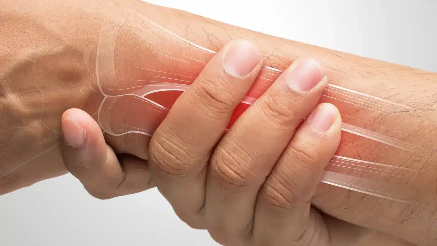
A broken wrist is a relatively common injury that can happen to anyone. This type of fracture typically occurs from direct impact to the wrist, such as a sports injury or a fall onto an outstretched hand. A broken wrist can significantly impact your daily activities and hinder your ability to use your hand correctly. Even after the initial treatment and healing process, proper care and rehabilitation are crucial for maintaining long-term wrist health. Fortunately, our whole-person approach is designed to help manage pain, support recovery, and improve long-term wrist function.

What Is A Distal Radius Fracture?

A distal radius fracture is a wrist injury that specifically affects the radius bone, which is one of two long bones in the forearm that run from the elbow to the wrist. The distal end of the radius is located at the wrist joint. It is responsible for connecting the hand to the forearm. There are eight small bones in the wrist joint, which allow the hand and arm to have a wide range of motion. A distal radius fracture can vary in severity, from a mild crack to a complete break through the bone.

A Comparison Between A Normal And Fractured Wrist

In a normal wrist, the radius and ulna bones work together to support the hand and allow for movement. The joint between these two bones is covered in cartilage, which helps reduce friction and allows for smooth movement. Ligaments, tendons, and muscles also play a crucial role in supporting the wrist joint.

In a fractured wrist, one or both of these bones may be broken, disrupting the proper alignment and stability of the joint. Such a fracture can lead to pain, swelling, and difficulty moving the hand and wrist.

Types Of Wrist Bone Fractures

  • Because the wrist consists of several small bones, there are different types of fractures that can occur. These include:

Scaphoid fracture: This is a fracture of the scaphoid bone, which is located on the side of the wrist leading up to the thumb. This type of fracture can affect the stability and function of the wrist joint due to its location and role in supporting the thumb.

  • Distal radius fracture: As noted above, this kind of fracture happens at the end of the radius bone near the wrist joint. It is one of the most common types of wrist fractures.
  • Chauffer’s (radial styloid) fracture: This fracture affects the radial styloid, which is a small bony bump on the thumb side of the wrist.

Barton’s fracture: This type of fracture occurs when the wrist joint is dislocated, causing the bones to break. It can have serious implications and may require surgery for proper treatment due to the complexity of the injury.

  • Ulnar styloid fracture: This fracture affects the ulnar styloid, which is a small bony bump on the pinky side of the wrist.

Pain And Other Symptoms Linked To Wrist Fractures

  • Wrist fractures often cause intense pain and discomfort, especially with movement. In addition to pain, other common symptoms of a broken wrist include:

Bruising: A bruise is a discoloration of the skin caused by damaged blood vessels leaking blood into the surrounding tissues. Bruising is a common symptom of a wrist fracture due to the impact or trauma that caused the injury.

Bone swelling: Bone swelling, also known as periosteal edema, is a buildup of fluid in the tissues surrounding the fracture site. This can cause visible swelling or a bump near the broken bone and may feel firm to the touch. Bone swelling is caused by inflammation in response to the fracture.

Inability to move the wrist: When the wrist is fractured, it may be difficult or impossible to move the hand and fingers normally. This is due to the pain, swelling, and instability caused by the fracture. In more severe cases, the wrist may become completely immobilized.

Why Do Wrist Fractures Occur?

  • Understanding the causes and risk factors of wrist fractures can help prevent future injuries. The most common causes of a wrist fracture include:

Age

As we age, our bones become more brittle and prone to fractures. This is because as we get older, our bodies produce less bone tissue, and the calcium levels in our bones decrease. This makes them weaker and more susceptible to breaks. As older individuals tend to be at a higher risk of falling, this further increases the risk of wrist fractures.

Gender

Women are more likely to experience wrist fractures, especially after menopause. This is the result of hormonal changes that can weaken bones by reducing bone density. Hormonal changes can also cause a lack of coordination and balance, putting women at a higher risk of falling.

Falls And Accidents

Falls, particularly onto an outstretched hand, are the leading cause of wrist fractures. Car accidents and other high-impact accidents can also result in wrist fractures.

Injuries

Injuries such as sports-related impacts or direct trauma to the wrist can cause fractures. For instance, a direct blow to the wrist while playing softball or football can result in a fracture.

In some cases, repetitive stress injuries may also contribute to a broken wrist. For example, a gymnast may experience fractures from repeatedly landing on their hands.

Medical Conditions

Certain medical conditions can weaken bones and increase the risk of fractures. It is essential to manage these conditions to prevent future injuries. The following medical conditions are linked to an increased risk of wrist fractures:

Osteoporosis

Osteoporosis is characterized by brittle and weak bones, making them more prone to fractures. It often affects older individuals, postmenopausal women, and those with a family history of the disease.

Osteogenesis Imperfecta

Osteogenesis imperfecta is also referred to as brittle bone disease. It’s a genetic disorder that causes bones to be more fragile and prone to fractures.

Cancer

Cancer can weaken bones and make them more susceptible to fractures. Certain cancer treatments, such as chemotherapy, may also increase the risk of bone fractures due to their impact on bone density.

Potential Risks Associated With An Untreated Broken Wrist

  • It is important to seek medical treatment for a broken wrist as soon as possible. Leaving a fractured wrist untreated can lead to potential risks and complications, including:
  • Malunion: When a bone heals in an incorrect position, it is called malunion. This can result in permanent deformity and reduced hand and wrist function.
  • Nonunion: Nonunion occurs when a bone fails to heal after an extended period of time. Nonunion can lead to chronic pain and decreased mobility.
  • Nerve damage: A broken wrist can lead to nerve damage, thereby resulting in symptoms such as tingling, numbness, and weakness in the hand and fingers.
  • Arthritis: If a fracture damages the cartilage or joint surfaces of the wrist, it can lead to arthritis. This can cause persistent pain and stiffness in the joint.

Compartment syndrome: In rare cases, a broken wrist can result in compartment syndrome, which is a serious condition caused by increased pressure within a muscle compartment. This can cut off blood flow and damage nerves and muscles. If left untreated, compartment syndrome can be life-threatening.

How Is Wrist Fracture Diagnosed?

If you’re experiencing any of the symptoms listed earlier, it is vital to seek medical attention immediately. A doctor will conduct a routine physical exam and may also order diagnostic tests to confirm a wrist fracture. These tests may include:

  • X-ray: This is the most common diagnostic test for bone fractures. X-rays create images of bones using low levels of radiation.
  • CT scan: A CT scan provides more detailed images than a regular X-ray. It can help doctors identify more complex fractures and determine their severity.
  • MRI: An MRI produces detailed images of bones and soft tissues by using powerful magnets and radio waves. This test is often used to examine complex fractures or to detect any nerve or ligament damage.
  • Bone scan: A bone scan involves injecting a tiny (and safe) amount of radioactive substance into the bloodstream. This allows doctors to detect any areas of increased bone activity, which can indicate a fracture.
  • Dual-energy X-ray absorptiometry (DXA): This test measures bone density and can help diagnose osteoporosis.

Common Treatment Options

  • Most traditional doctors will recommend more conventional methods for treating wrist fractures. Some of the more common treatment options for a fractured wrist include the following:

Medications

If you’re experiencing pain or swelling, over-the-counter (OTC) pain relievers like acetaminophen or ibuprofen can provide relief. In more severe cases, your doctor may prescribe a stronger pain medication. However, pain medications—especially prescription medications—can have side effects and, in some cases, carry a risk of dependence. They help manage pain but do not directly heal the fracture.

Immobilization

Immobilizing the wrist can help alleviate pain and prevent further injury. This is achieved through splinting or casting, which keeps the bones in proper alignment while they heal.

Physical Therapy

Physical therapy can help regain mobility, flexibility, and strength in the wrist after a fracture has healed. A physical therapist may also recommend exercises to improve grip strength and prevent future injuries.

Surgery

Surgery is typically reserved for more severe fractures or cases where the bones are not aligned properly. Surgery is incredibly invasive and can be risky as well since it involves cutting the skin and using screws, pins, or plates to hold the bones in place. It could potentially lead to complications or infection.

  • That said, the following are the two types of surgery used to treat wrist fractures:

Closed reduction: This involves manipulating the bones back into place without making any incisions. It’s typically performed under local or general anesthesia and can be done in an outpatient setting. Although not as invasive, there’s still a risk of nerve damage or further bone damage if the bones are not aligned properly.

Open reduction: This type of surgery involves making an incision to realign the bones and then using plates, screws, or pins to hold them in place. It’s a more invasive approach that may require a hospital stay and carries a higher risk of infection or nerve damage. Additionally, recovery time for this surgery is longer compared to closed reduction.

Can Broken Bones Heal Faster With Conventional Treatment?

While conventional treatments for wrist fractures can be effective in most cases, the bones can take a considerable amount of time to fully heal. This is due to the natural healing process of bones, which can take anywhere from six to eight weeks or longer, depending on the severity of the fracture.

Some conventional treatments, particularly surgery, are invasive and carry risks such as infection, stiffness, and a longer rehabilitation period when they are necessary.

We offer an alternative treatment plan that is non-invasive, non-surgical, and drug-free. Our treatment protocol can help expedite the healing process and provide relief from pain and discomfort.

If you’ve already undergone surgery to repair a wrist fracture, our Relatyv Neurofunctional Pain Management approach can still be beneficial by reducing inflammation and chronic pain that may have developed after the surgery, or that may have continued to linger post-surgery.

Our Relatyv Neurofunctional Pain Management Approach for Wrist Fracture Recovery

We use a Neurofunctional Pain Management approach to help patients with wrist fractures. Essentially, we use several treatment modalities (all of which are drug-free and non-invasive) to target the neurological causes of pain and promote accelerated healing.

Support Your Recovery and Restore Wrist Function

Our goal is to provide a personalized treatment plan that helps reduce pain, improve mobility, and support your recovery using non-invasive therapies that complement your overall treatment plan.

Our experienced team is here to help you manage pain, regain function, and recover with confidence. Contact us to learn how our Relatyv Neurofunctional Pain Management approach may support your recovery.

Relatyv - RLS

Restless Leg Syndrome (RLS)

Restless Leg Syndrome (RLS) 1280 720 Relatyv

Restless Leg Syndrome

Living with restless leg syndrome (RLS) can be challenging and disruptive to daily life. It is a neurological condition that can make it challenging to fall asleep or stay asleep, which can have a significant impact on your quality of life. For example, it can lead to daytime drowsiness, irritability, and difficulty concentrating, all of which can affect work performance and personal relationships. Fortunately, there are several ways that you can manage the symptoms and improve your overall well-being.

Our Relatyv Neurofunctional Pain Management approach uses non-invasive therapies designed to help reduce discomfort, improve function, and support overall nervous system health for people living with restless leg syndrome.

What Is Restless Leg Syndrome?

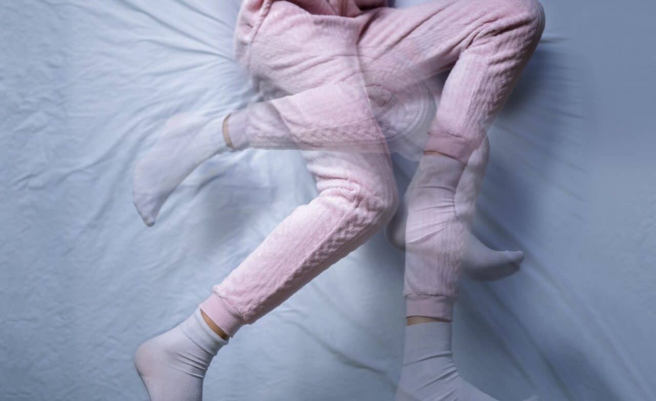
Restless leg syndrome is a common, yet often misunderstood, condition that affects the nervous system. It is classified as a neurological disorder, due to its impact on the body’s nerve signals and functions. RLS is characterized by the urge to move one’s legs, usually accompanied by uncomfortable sensations in the lower limbs.

Who Experiences Restless Legs Syndrome

RLS affects approximately 7 to 11% of the Western population. While anyone can develop RLS, it is more commonly seen in women, older adults, and those with a family history of the condition. It’s also important to note that about two-thirds of those affected experience RLS as the primary condition, while the remaining third may experience it as a symptom of an underlying condition.

How It Affects The Body

Aside from the physical discomfort, RLS can also have an emotional impact on those who experience it. The constant urge to move can cause frustration, irritability, and difficulty sleeping. Lack of sleep and constantly disrupted rest can lead to fatigue, decreased concentration, and even depression in some cases.

Recognizing The Signs And Symptoms

  • RLS can present differently for everyone, but common symptoms include:

Leg Twitching

Some individuals may experience involuntary leg movements, such as twitching or jerking, during the night or while trying to fall asleep.

Urge To Move

The primary symptom of RLS is an uncontrollable urge to move the legs. These movements are often described as a crawling or creeping sensation in the legs, and they typically occur when you’re lying down or sitting for long periods.

Tingling And Burning Sensations In Legs

It is also common for individuals with RLS to experience tingling, burning, or itching sensations in their legs. Such sensations can be mild or severe and can produce discomfort and restlessness.

Pain And Discomfort In Arms Or Legs

While the name suggests that RLS only affects the legs, some individuals may also experience symptoms in their arms. These sensations can be just as uncomfortable and disruptive to daily life.

Back Pain

In some cases, RLS can also cause lower back pain, making it difficult for individuals to find relief by changing their position or moving their legs.

Urge To Scratch Legs

RLS can also lead to an urge to scratch or rub the legs. Constant scratching can damage the skin and lead to further discomfort.

Causes Of Restless Leg Syndrome

  • The cause of RLS is still unknown. However, researchers have identified several factors that could contribute to its development. These include:

Genetic Factors

RLS is believed to have a genetic component, as it tends to run in families. Studies have identified specific gene variations that may increase the risk of developing RLS.

Neurological Abnormalities

RLS is thought to be related to abnormalities of the nervous system, which disrupt the normal signaling and processing of sensations. For example, individuals with RLS may have lower levels of dopamine. Dopamine is a chemical in your brain that helps with controlling muscle movement.

Kidney Failure

In rare cases, RLS has been linked to kidney failure. This is because individuals with kidney disease may have an imbalance of iron and other minerals in their blood, which can contribute to RLS symptoms.

Iron Deficiency Anemia

Iron is essential for producing dopamine and plays a role in nerve signaling. Therefore, individuals with iron deficiency anemia may be more likely to develop RLS.

Deficiency In Cortisol

Cortisol is an important hormone in the body that is meant to help regulate stress and inflammation. Low levels of cortisol have been linked to RLS symptoms, as it may impact nerve signaling and muscle movement.

Factors That Heighten The Risk Of Developing RLS

  • There are numerous risk factors that could increase the likelihood of developing RLS, including:

Genetic Background

As mentioned earlier, having a family history of RLS can increase the risk of developing the condition.

Attention Deficit Hyperactivity Disorder (ADHD)

ADHD is a neurodevelopmental disorder that affects your ability to focus, control impulsive behaviors, and regulate emotions. It’s been shown that there is a connection between ADHD and RLS, with ADHD being an independent risk factor for developing RLS, as both conditions have been linked to abnormalities in dopamine levels.

Pregnancy

Pregnant women often experience RLS symptoms, especially in the third trimester. This may be due to hormonal changes or increased pressure on the legs.

Dialysis

Dialysis is a medical treatment used to filter waste and excess fluid from the blood when the kidneys are no longer able to function properly. Individuals undergoing dialysis may have an increased risk of developing RLS due to imbalances in iron and other minerals in their blood.

Anxiety Disorders

Anxiety disorders are mental health conditions characterized by excessive worry, fear, and tension. Anxiety can disrupt sleep and contribute to RLS symptoms due to increased muscle tension and stress on the nervous system.

Medications

Certain medications, including antihistamines and antidepressants, can worsen RLS symptoms or trigger them in individuals who are at risk for developing the condition. This is because such medications can impact dopamine levels and nerve signaling.

Medical Conditions

  • Several medical conditions may increase the risk of RLS, including:

Osteoarthritis: This condition is known to cause chronic joint stiffness and pain. These symptoms make it difficult for individuals to find comfortable positions and may lead to RLS symptoms. Additionally, medications used to treat osteoarthritis can also worsen the symptoms of RLS.

Rheumatoid arthritis: Similar to osteoarthritis, rheumatoid arthritis can cause joint pain and stiffness that may contribute to RLS symptoms due to difficulty finding comfortable positions and potential medication side effects.

Peripheral neuropathy: This condition involves nerve damage and can cause sensations of tingling, burning, or pain in the legs. It is estimated that up to one-third of individuals with peripheral neuropathy also experience RLS symptoms.

  • Fibromyalgia: This condition can result in chronic pain and fatigue, and it has been linked to RLS symptoms in some individuals due to increased muscle tension and nerve signaling disruption.

Diagnosing Restless Leg Syndrome

  • In addition to asking about your medical history and symptoms, a doctor will perform the following to help diagnose RLS:

Neurological exam: A neurological exam is a series of tests to assess the function and health of your nervous system (which includes the evaluation of your brain, spinal cord, and nerves). This can help doctors rule out other potential causes of RLS symptoms.

  • Physical exam: A physical exam will be done to check for any underlying medical conditions contributing to your RLS symptoms. For example, a doctor may examine your skin and reflexes for signs of anemia or nerve damage.

Blood tests: Blood tests can be ordered to help identify any potential underlying medical conditions or deficiencies that may be causing RLS symptoms. These tests may include checking on iron levels, thyroid function, and kidney function.

How Restless Legs Syndrome Is Commonly Managed

  • RLS is a relatively common condition, which means that various management strategies are available. Some of the most common approaches recommended by more conventional doctors include:

Practicing Good Sleep Hygiene

Getting enough sleep and ensuring good sleep quality are essential for managing RLS symptoms. This includes going to sleep on a regular schedule, creating a sleeping environment that is comfortable, and avoiding activities that disrupt sleep. Doing so can help reduce the frequency and severity of RLS symptoms since getting enough rest can help regulate dopamine levels and decrease stress on the nervous system.

Engaging In Physical Activity

Regular physical activity can help to improve RLS symptoms by promoting better sleep and reducing stress. Additionally, exercise can help increase dopamine levels and promote overall nerve health by stimulating blood flow to the nerves. However, it is important to avoid exercising too close to bedtime as this may worsen RLS symptoms by increasing body temperature and adrenaline levels.

Minimizing Alcohol And Caffeine Intake

Both alcohol and caffeine have been linked to disrupted sleep patterns and may worsen RLS symptoms in some individuals. Both substances can impact dopamine levels and brain activity, increasing nerve signaling and muscle tension. Limiting or avoiding consuming these substances, especially close to bedtime, is recommended.

Applying Heat Or Ice Pack

Applying heat or ice packs to the legs can help relieve discomfort and reduce RLS symptoms. Heat can relax tense muscles, while cold can numb nerve endings and provide temporary relief from sensations.

Ensuring Adequate Intake Of Iron, Folate, And Magnesium

Deficiencies in iron, folate, and magnesium have been linked to RLS symptoms. Therefore, ensuring adequate intake of these minerals through a balanced diet or supplements is critical. However, consulting with a doctor before taking any supplements is crucial to avoid potential interactions with other medications.

Conventional Treatment Options

  • If the previously mentioned approaches are not effective, a doctor may recommend the following conventional treatment options for RLS:

Medications

There are many types of medications and supplements that can help alleviate RLS symptoms. However, keep in mind that many medications can cause various side effects. In addition, most medicines will only mask the symptoms and won’t address the underlying causes of your symptoms. That being said, the following medication options may be recommended to help manage RLS symptoms:

Non-steroidal anti-inflammatory drugs: These medications can help reduce inflammation and leg pain that may contribute to RLS symptoms. However, they should be used cautiously, as long-term use can lead to gastrointestinal issues.

  • Iron supplements: If blood tests show an iron deficiency, your doctor will likely recommend iron supplements to help increase your iron levels and help improve RLS symptoms.
  • Dopaminergic agents: These medications can help regulate dopamine levels and reduce RLS symptoms. However, they may also cause side effects such as nausea, dizziness, and headache.
  • Benzodiazepines: These sedative medications can help improve sleep and reduce muscle tension, leading to better management of RLS symptoms. However, they may also cause addiction and withdrawal symptoms.
  • Anticonvulsants: These medications can help reduce nerve activity and promote relaxation, leading to improved RLS symptoms. However, they may also cause side effects such as drowsiness and dizziness.
  • Opioids: In severe cases, a doctor may prescribe opioids to help manage RLS symptoms. However, these medications can be very addictive and should be used cautiously.

Cognitive-Behavioral Therapy (CBT)

CBT is a type of therapy that can help individuals identify and change thought patterns and behaviors contributing to their RLS symptoms. For example, a therapist may help someone reduce stress and anxiety by teaching relaxation techniques or creating a sleep plan to improve rest. This approach can help reduce stress levels, improve sleep, and manage RLS symptoms.

Pneumatic Pressure Therapy

Pneumatic pressure therapy involves wearing inflatable leg wraps that apply pressure to the legs, promoting better circulation and reducing RLS symptoms. This method can provide temporary relief from discomfort and help improve sleep quality.

Deep Brain Stimulation (DBS)

DBS is a surgical procedure involving the application of electrodes into specific areas of the brain to help regulate nerve activity and reduce RLS symptoms. This treatment option is only recommended in severe cases where other methods have been unsuccessful.

Living With Restless Leg Syndrome

While RLS is a chronic condition, it can be effectively managed with the proper treatment plan. Working closely with your doctor to find the best management strategies for your individual needs is essential.

How Relatyv Neurofunctional Pain Management Tackle Restless Leg Syndrome Pain

At Relatyv, we’ve developed a protocol that takes a whole-person, Neurofunctional Pain Management approach to treating pain and inflammation caused by conditions such as RLS. Our specialists focus on helping manage the neurological factors that may contribute to your symptoms while complementing your overall treatment plan. For example, we use electroanalgesia to stimulate nerve pathways, increasing blood circulation and reducing pain signals to the affected area. This approach is designed to help reduce discomfort, support nerve function, and improve quality of life.

Our team may also recommend dietary changes, IV therapy, and other non-invasive techniques to help manage your RLS symptoms effectively. Through our personalized treatment plans, we aim to improve your quality of life by addressing the underlying causes of your condition using drug-free, non-invasive, non-chiropractic, and non-surgical methods.

End Restless Leg Syndrome Pain

Restless leg syndrome can significantly impact an individual’s quality of life, but it doesn’t have to be a lifelong struggle. The proper treatment plan and lifestyle changes can help manage RLS symptoms. We take a unique approach to treating RLS by addressing the underlying neurological causes of your symptoms.

Our goal is to help reduce pain, improve sleep and daily function, and support long-term well-being through personalized, non-invasive care. Contact us to learn how our Relatyv Neurofunctional Pain Management approach may complement your treatment plan.

Relatyv - Radial Tunnel Syndrome

Radial Tunnel Syndrome Pain

Radial Tunnel Syndrome Pain 2000 1127 Relatyv

Radial Tunnel Syndrome

Radial tunnel syndrome (RTS) is a relatively uncommon condition that affects the forearm and wrist. The annual incident rate of RTS is estimated to be only 0.03%. However, RTS can significantly impact a person’s daily life by limiting their ability to perform simple tasks such as gripping objects, typing, and even writing. It can also cause significant discomfort and affect one’s quality of life if left untreated. Fortunately, at Relatyv Neurofunctional Pain Management, we offer an effective pain management solution to help those suffering from RTS.

What Is Radial Tunnel Syndrome?

RTS is a condition that affects the radial nerve, which runs from the neck down to the hand. The radial nerve controls movements of the triceps muscle in the upper arm. Also, it helps with sensation on the back of the hand.

When this nerve is irritated or becomes compressed, it can cause weakness, numbness, and pain in certain areas of the arm and hand. RTS is often compared to “tennis elbow” because it can cause similar symptoms, although it affects a different part of the arm.

Where Is The Radial Tunnel Located?

The radial tunnel is located on the lateral (outer) side of the elbow, between the humerus bone and the supinator muscle. It comprises various structures, including the radial nerve, posterior interosseous nerve, brachioradialis muscle, and extensor carpi radialis longus muscle.

The supinator muscle wraps around the upper part of the radius bone. It plays a role in supination (rotation of the forearm). The extensor carpi radialis longus muscle helps with wrist extension, while the brachioradialis muscle flexes the elbow. All of these structures interact with the radial nerve within the radial tunnel.

Common Symptoms Associated With Radial Tunnel Syndrome

  • Because the radial nerve controls both muscle movements and sensation, RTS can cause various symptoms. These may include:
  • Aching or burning pain in the forearm: RTS pain is typically felt in the outer part of the forearm, closest to the elbow. This can sometimes radiate down to the wrist and hand.
  • Aggravation with certain movements: Activities that involve repetitive wrist movements or gripping can exacerbate RTS pain. This includes tasks like typing, using a screwdriver, or playing sports such as tennis.
  • Painful grip: People with RTS may experience pain when trying to grip objects, making it difficult to perform simple everyday tasks, such as holding a coffee cup or a phone.

What Are The Most Probable Causes Of The Condition?

  • Knowing the potential causes of radial tunnel syndrome can help you prevent future occurrences and choose the most effective treatment options. The most common causes of RTS include:

Repetitive Wrist And Forearm Movements

Repetitive wrist and forearm movements, especially those involving twisting or gripping, can strain the muscles and tendons in the arm. This strain can lead to inflammation and compression of the radial nerve within the tunnel. Activities that may contribute to RTS in this way include playing certain sports, using power tools, or typing on a computer for extended periods.

Trauma Or Injury

A fall, impact, or other direct trauma to the elbow can cause damage to the structures within the radial tunnel and lead to RTS. This may include fractures or dislocations of the humerus bone or injuries to surrounding muscles and tendons. There are countless ways that someone can experience physical trauma to the elbow, so it’s essential to be aware of potential harm when participating in risky activities. For example, falling off a bike or being in a car accident could lead to damage in the radial tunnel.

Underlying Medical Conditions

  • Some underlying conditions can increase your risk of developing radial tunnel syndrome. These include the following:
  • Ligament injuries: Injuries to the ligaments in the elbow, such as sprains or tears, can put pressure on the radial nerve, thereby causing compression.

Tumors or ganglion cysts: Tumors and ganglion cysts are abnormal growths that can sometimes develop within the elbow joint or along the path of the radial nerve. These masses can compress the nerve and cause symptoms if they grow large enough.

Diabetes: There is a higher risk of developing nerve damage, including compression of the radial nerve, if you have diabetes. This is because the small blood vessels that feed and nourish your nerves are more prone to damage if you have high blood sugar levels.

Osteoarthritis: Osteoarthritis is a degenerative joint disease that can affect the elbow joint. The condition causes cartilage to wear away. This, in turn, results in your bones beginning to rub against each other, which can lead to the development of bone spurs. Bone spurs can compress surrounding structures such as muscles and nerves.

Gout: Gout is an inflammatory arthritis that can cause swelling and joint pain. It tends to affect the big toe but can also develop in other joints, such as the elbow. Excess uric acid crystals can build up and create deposits in the joint and may compress nerves.

  • Rheumatoid arthritis: Rheumatoid arthritis is an autoimmune disorder known for causing joint inflammation. This inflammation can lead to swelling and stiffness, which can compress nerves and cause symptoms.

When To Seek Medical Attention

You should seek medical attention if your symptoms worsen or start affecting daily activities. A doctor will physically examine your arm and order diagnostic tests to assess the potential cause of your symptoms. These diagnostic tests can include imaging studies such as X-rays, MRI, or nerve conduction tests. It is also vital to seek medical attention if there are any signs of infection around the elbow joint, such as redness, warmth, or swelling.

How Is Radial Tunnel Pain Diagnosed And Evaluated?

  • To diagnose radial tunnel syndrome, a doctor will typically perform the following steps:
  • Evaluation of medical history: The doctor will inquire about any previous injuries or medical conditions that may be contributing to the symptoms.
  • Clinical examination: A physical exam will be conducted to check for any tenderness or weakness in the wrist and hand, as well as any decreased range of motion.
  • Nerve conduction study: This test involves measuring the speed and strength of electrical signals sent through the nerves in the arm. It can help determine if there is any nerve damage or compression.
  • Imaging tests: X-rays, MRI, or ultrasound may be ordered to visualize the structures in and around the elbow joint and identify any potential causes of nerve compression.
  • MRI: An MRI will provide the doctor with images of the soft tissues in and around the elbow joint, making it useful for detecting abnormalities such as cysts or tumors.
  • Ultrasound: Ultrasound uses high-frequency sound waves to develop images of the body’s structures. It can help identify any swelling or inflammation around the radial nerve.

Differential Diagnosis Of Radial Tunnel Pain

A differential diagnosis is a list of possible conditions that could be causing the symptoms someone is experiencing. Several conditions can cause symptoms similar to radial tunnel syndrome, which is why it’s essential to rule out other potential causes. Some possible differential diagnoses for RTS may include:

Cubital tunnel syndrome: Cubital tunnel syndrome is when the ulnar nerve is compressed at your elbow. This condition can lead to feelings of numbness and pain in your hand and fingers. It can sometimes be mistaken for RTS, but the area of compression is different.

  • Tennis elbow: Tennis elbow is when inflammation and pain develop in the tendons on the outside of the elbow. It can cause similar symptoms to RTS, but the underlying cause is different.

Wrist tendinitis: This refers to inflammation and irritation of the wrist tendons. This can cause hand pain and weakness. Wrist tendinitis can sometimes be mistaken for RTS because it may also involve symptoms in the hand and fingers.

Thoracic outlet syndrome: This condition involves compression of nerves or blood vessels in the space between the collarbone and first rib, causing pain and tingling sensations in the arm. It can be misdiagnosed as RTS if the symptoms primarily affect the forearm and hand.

What Treatment Options Are Available?

Traditional treatment for radial tunnel syndrome may include a combination of conservative measures and more invasive procedures. Conventional doctors will recommend the most appropriate treatment based on their diagnosis of the underlying cause of RTS, as well as the severity and duration of symptoms.

Conservative Measures & Medications

  • The following conservative measures are often used to manage RTS symptoms:
  • Rest: Simply taking a break from physical activities that strain the forearm muscles and tendons can help alleviate symptoms by allowing the affected area to heal.
  • Ergonomic modifications: Making changes to work or home environments, such as using a wrist brace or modifying computer equipment, can help reduce strain on the arm and alleviate symptoms.

Physical therapy: A physical therapist can prescribe exercises and stretches tailored to your condition that will help improve your arm’s flexibility and strength. Doing so can relieve pressure on the radial nerve and improve symptoms.

  • NSAIDs: NSAIDs, or non-steroidal anti-inflammatory drugs, are commonly recommended to provide temporary relief from pain and inflammation associated with RTS.
  • Oral steroids: In some cases, a doctor may prescribe oral steroids to reduce inflammation and alleviate symptoms.
  • Topical analgesics: Creams or patches containing analgesic medication may also provide short-term pain relief.

Surgery

In severe cases of RTS or when conservative measures have been unsuccessful, surgery may be recommended. The type of surgical procedure that is recommended will depend on the underlying cause of RTS. For example, if there is a cyst or tumor causing compression of the radial nerve, it may need to be removed surgically. Surgery can also involve releasing tight structures or cutting muscles to relieve pressure on the nerve.

All surgical procedures involve potential risks and complications that should be considered. For example, surgery can sometimes lead to further nerve damage or infection. That being said, the following procedures are commonly used to treat RTS:

  • Release surgery: Release surgery involves releasing any tight structures or cutting muscles to relieve pressure on the radial nerve. It is often performed arthroscopically, using small incisions and specialized tools.

Decompression surgery: If a cyst or tumor is causing compression on the radial nerve, surgery may be required to remove it and relieve pressure on the nerve. Decompression surgery can also remove any scar tissue or adhesions that may be compressing the nerve.

  • Nerve transposition: In cases where the nerve is severely compressed and damaged, moving the nerve to a different location in the arm may be necessary. This procedure is known as a nerve transposition.

The Relatyv Neurofunctional Pain Management Approach To Radial Tunnel Syndrome Pain

Our whole-person approach combines drug-free, non-invasive therapies designed to help manage pain, improve function, and support your overall recovery. These treatments are intended to complement your broader medical care.

Seek Out Effective Radial Tunnel Pain Relief

Radial tunnel syndrome can be a debilitating and frustrating condition, but it is important to remember that there are effective treatment options available to help manage your pain. A correct diagnosis is vital to determine the best course of action for managing RTS pain.

Our Relatyv Neurofunctional Pain Management approach focuses on helping reduce pain, improve mobility, and support long-term function through personalized, non-invasive care. Contact us to learn how our treatment approach may complement your recovery plan.

Relatyv - Plantar Fasciitis

Plantar Fasciitis

Plantar Fasciitis 1920 1082 Relatyv

Pain on the Bottom of Your Foot? Understanding Plantar Fasciitis

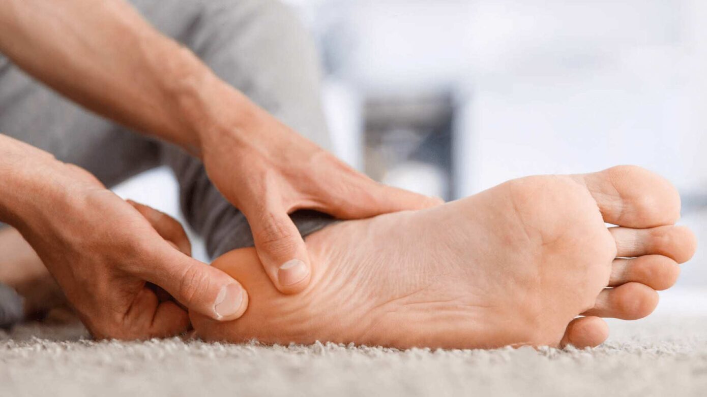
Pain on the bottom of your foot can make everyday activities like walking, standing, and exercising difficult. One of the most common causes of this type of pain is plantar fasciitis. Understanding the condition, its causes, symptoms, and treatment options can help you find relief and prevent it from becoming a long-term problem.

What Is Plantar Fasciitis?

Plantar fasciitis is one of the most common causes of heel pain. It develops when the plantar fascia—a thick band of connective tissue that runs from the heel bone to the toes—becomes irritated or overloaded, leading to pain and stiffness along the bottom of the foot. While inflammation may be present early on, long-standing plantar fasciitis often involves degeneration of the tissue rather than ongoing inflammation.

Plantar fasciitis affects millions of people and is estimated to occur in approximately 10% of the population at some point during their lifetime.

Causes of Plantar Fasciitis

Damage or repetitive stress to the plantar fascia can lead to pain and irritation. While plantar fasciitis most commonly affects one foot, it can occur in both feet.

Standing or Walking for Long Periods

Prolonged standing or repetitive walking places additional stress on the plantar fascia. Jobs that require extended periods on your feet—such as nursing, food service, retail, or manufacturing—can increase your risk.

People who regularly run, hike, participate in high-impact sports, or train for endurance events may also be more likely to develop plantar fasciitis.

Wearing Shoes Without Proper Support

Shoes that lack adequate arch support or cushioning can increase strain on the plantar fascia. Thin-soled shoes, flip-flops, and high heels may contribute to the development of plantar fasciitis, especially if you’re standing or walking for extended periods.

Flat Feet or High Arches

Both flat feet and high arches can alter how weight is distributed across the foot, increasing stress on the plantar fascia and making injury more likely.

Exercising Without Warming Up

Stretching and warming up before exercise helps prepare the muscles and connective tissues for activity. Tight calf muscles or poor flexibility can increase stress on the plantar fascia and raise the risk of injury.

Symptoms of Plantar Fasciitis

Although heel pain can have many causes, plantar fasciitis has several characteristic symptoms. If you experience any of the following, it’s important to consult a healthcare provider for an accurate diagnosis.

  • Sharp pain under the heel or arch of the foot
  • Pain that’s often worse with the first steps in the morning
  • Pain after standing or walking for long periods
  • Difficulty walking or exercising comfortably
  • Increasing pain if the condition is left untreated

Sharp Heel or Arch Pain

The hallmark symptom of plantar fasciitis is a sharp pain in the heel or arch. It is often most noticeable when taking your first steps after waking up or after sitting for an extended period.

Pain That Gradually Worsens

Symptoms may begin as a mild ache and gradually worsen over time if repetitive stress continues. Pain often becomes more noticeable as the plantar fascia has less opportunity to recover between periods of activity.

Difficulty Walking

As pain increases, everyday activities such as walking, running, climbing stairs, or exercising may become increasingly difficult.

Treatment Options for Plantar Fasciitis

Treatment for plantar fasciitis typically begins with conservative measures designed to reduce pain, improve flexibility, and allow the plantar fascia to heal. Surgery is generally reserved for cases that do not improve with non-surgical treatment.

Rest

Reducing activities that place excessive stress on the foot allows the plantar fascia time to recover. Limiting prolonged standing and avoiding high-impact activities may help relieve symptoms.

Stretching Exercises

Stretching the calf muscles, Achilles tendon, and plantar fascia can improve flexibility, reduce tension, and help relieve heel pain.

Supportive Footwear

Wearing supportive shoes with proper arch support and cushioning can reduce stress on the plantar fascia and improve comfort throughout the day.

Corticosteroid Injections

Corticosteroid injections may provide temporary pain relief for some people but are generally used selectively because repeated injections can increase the risk of weakening or damaging the plantar fascia.

Surgery

Surgery is typically considered only after conservative treatments have failed.

Common procedures include:

  • Gastrocnemius recession, which lengthens the calf muscle to reduce stress on the plantar fascia.
  • Plantar fascia release, which reduces tension by making small incisions in the fascia.

Although surgery can benefit carefully selected patients, it carries the same risks associated with any surgical procedure, including infection, nerve injury, scarring, prolonged recovery, and persistent pain.

Our Relatyv Neurofunctional Pain Management Approach for Plantar Fasciitis

At Relatyv, our Neurofunctional Pain Management approach combines drug-free, non-invasive therapies designed to help reduce pain, improve mobility, and support the body’s natural healing process.

Treatment may include electroanalgesia, a non-invasive therapy that uses electrical stimulation to help manage pain and encourage normal nerve signaling. Electroanalgesia has been used for decades as part of comprehensive pain management programs.

Treatment plans are individualized based on each patient’s condition, symptoms, and response to care. When clinically appropriate, care may also include IV therapy, nutritional support, and lifestyle recommendations to complement your overall treatment plan.

Find Relief from Plantar Fasciitis Pain

Plantar fasciitis can make even simple daily activities uncomfortable, but effective treatment options are available.

At Relatyv, we provide personalized, non-invasive care designed to help reduce pain, improve function, and support long-term foot health. Our comprehensive approach may include electroanalgesia, IV therapy when appropriate, nutritional support, lifestyle guidance, and patient education to complement your overall treatment plan and help you move more comfortably. Contact us to learn how our Neurofunctional Pain Management approach may support your recovery.
Realtyv - Interstitial Cystitis

Interstitial Cystitis

Interstitial Cystitis 2560 1442 Relatyv

Understanding Interstitial Cystitis (IC)

Interstitial cystitis (IC), also known as painful bladder syndrome, is a chronic condition that affects the bladder and causes persistent pelvic pain, pressure, and urinary symptoms. These symptoms can interfere with daily activities, work, sleep, relationships, and overall quality of life.

This guide explains the anatomy of the bladder, the causes and symptoms of IC, how it’s diagnosed, and the treatment options that may help manage discomfort. We’ll also discuss how our Relatyv Neurofunctional Pain Management approach may complement your overall treatment plan.

Understanding the Urinary System

The urinary system consists of four primary structures:

  • Kidneys
  • Ureters
  • Bladder
  • Urethra

The kidneys filter waste products and excess fluid from the bloodstream to produce urine. Urine travels through the ureters into the bladder, where it is stored until it is released through the urethra.

The bladder is a muscular organ located in the lower abdomen. As it fills, nerves within the bladder communicate with the brain to signal when it’s time to urinate. A healthy bladder typically stores approximately 400–600 milliliters of urine before emptying.

In addition to removing waste, the urinary system helps regulate fluid balance, electrolyte levels, blood pressure, and red blood cell production.

Conditions such as interstitial cystitis can disrupt normal bladder function and lead to ongoing pain and urinary symptoms.

What Is Interstitial Cystitis?

Interstitial cystitis (IC) is a chronic bladder pain condition characterized by persistent bladder discomfort lasting six weeks or longer without an identifiable infection or other obvious cause. It is also commonly referred to as painful bladder syndrome.

Although the exact cause remains unknown, IC is thought to involve irritation of the bladder lining, altered nerve signaling, inflammation, and dysfunction of the bladder wall. Over time, these changes may reduce bladder capacity and contribute to chronic pelvic pain and urinary urgency.

Symptoms of Interstitial Cystitis

Symptoms vary from person to person and may fluctuate over time. Some individuals experience occasional flare-ups, while others have persistent symptoms.

Pelvic Pain and Pressure

Persistent pain or pressure in the bladder, lower abdomen, pelvis, or perineum is one of the hallmark symptoms of IC. Symptoms often worsen as the bladder fills and improve after urination.

Urinary Urgency and Frequency

People with IC often feel a frequent or urgent need to urinate, even when the bladder contains very little urine. Changes in bladder sensitivity and reduced bladder capacity may contribute to these symptoms.

Urinary Incontinence

Although less common, some individuals experience urinary leakage, particularly during periods of increased urgency.

Pain During Intimacy

Pelvic pain and muscle tension associated with IC can make sexual activity uncomfortable or painful for some individuals.

Possible Causes of Interstitial Cystitis

The exact cause of IC remains unknown, but researchers believe several factors may contribute to its development.

Bladder Lining Dysfunction

Damage to the protective lining of the bladder may allow substances in the urine to irritate the bladder wall, leading to pain, inflammation, and increased sensitivity.

Pelvic Floor Dysfunction

Tight or dysfunctional pelvic floor muscles may contribute to pelvic pain, urinary urgency, and bladder symptoms. Pelvic floor physical therapy is often included in treatment plans.

Autoimmune Conditions

Some studies have identified an association between IC and autoimmune disorders such as fibromyalgia, irritable bowel syndrome (IBS), and chronic fatigue syndrome. While the relationship is not fully understood, immune system dysfunction may play a role in some individuals.

Nerve Dysfunction

Changes in how nerves communicate between the bladder, spinal cord, and brain may contribute to heightened pain sensitivity and abnormal bladder signaling.

How Interstitial Cystitis Can Affect Daily Life

Living with IC often extends beyond physical discomfort. Persistent symptoms may affect many aspects of daily life.

Physical Limitations

Chronic pain, urinary urgency, and frequent bathroom visits can make exercise, travel, work, and social activities more challenging.

Emotional Well-Being

Managing ongoing symptoms may contribute to increased stress, anxiety, depression, and reduced quality of life.

Work and Productivity

Frequent interruptions, pain flare-ups, and fatigue may affect concentration, attendance, and productivity in the workplace.

How Is Interstitial Cystitis Diagnosed?

Because there is no single test that confirms IC, diagnosis typically involves ruling out other possible causes of symptoms.

Your healthcare provider may recommend:

  • Medical history review
  • Physical examination
  • Urine and blood testing
  • Urodynamic testing
  • Cystoscopy
  • Ultrasound or MRI when appropriate

An accurate diagnosis helps guide the most appropriate treatment plan.

Conventional Treatment Options

Treatment for interstitial cystitis is highly individualized. Most treatment plans combine multiple approaches to help manage symptoms.

Medications

  • Tricyclic antidepressants may help reduce pain and bladder sensitivity.
  • Antihistamines may reduce bladder irritation in some patients.
  • Pain relievers, including NSAIDs or acetaminophen, may help manage discomfort. Prescription medications may be considered for more severe symptoms.

Because medications affect everyone differently, treatment decisions should be made with your healthcare provider after discussing potential benefits and risks.

Complementary Therapies

Some individuals benefit from combining medical treatment with complementary therapies.

Bladder Training

Structured bladder training programs can help improve bladder capacity and reduce urinary urgency over time.

Pelvic Floor Therapy

Trigger point therapy and pelvic floor physical therapy may help reduce muscle tension and improve pelvic pain.

Bladder Instillation

Bladder instillation introduces medication directly into the bladder through a catheter to help reduce irritation and improve symptoms in some patients.

Our Relatyv Neurofunctional Pain Management Approach for Interstitial Cystitis

At Relatyv, our Neurofunctional Pain Management approach combines drug-free, non-invasive therapies designed to help reduce pain, improve function, and support overall nervous system health.

Treatment plans are personalized and may include electroanalgesia, IV therapy when clinically appropriate, nutritional support, and lifestyle recommendations. Our goal is to complement your overall treatment plan while helping improve comfort and daily function.

Find Relief from Interstitial Cystitis Symptoms

Living with interstitial cystitis can be physically and emotionally challenging, but effective management strategies are available.

At Relatyv, we provide personalized, non-invasive care designed to help reduce pain, improve bladder comfort, and support long-term quality of life. If you’re looking for additional options to complement your current treatment plan, our Neurofunctional Pain Management approach may help you better manage chronic symptoms and return to the activities that matter most.

Back to top
Privacy Preferences

When you visit our website, it may store information through your browser from specific services, usually in the form of cookies. Here you can change your Privacy preferences. It is worth noting that blocking some types of cookies may impact your experience on our website and the services we are able to offer.

Click to enable/disable Google Analytics tracking code.
Click to enable/disable Google Fonts.
Click to enable/disable Google Maps.
Click to enable/disable video embeds.
Our website uses cookies, mainly from 3rd party services. Define your Privacy Preferences and/or agree to our use of cookies.